. 2014 Feb 26;34(9):3183-92.
doi: 10.1523/JNEUROSCI.4958-13.2014.

Glutamate and dopamine transmission from midbrain dopamine neurons share similar release properties but are differentially affected by cocaine

Affiliations

Glutamate and dopamine transmission from midbrain dopamine neurons share similar release properties but are differentially affected by cocaine

Martín F Adrover et al. J Neurosci. .

Abstract

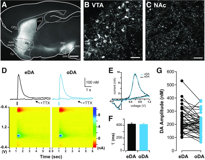
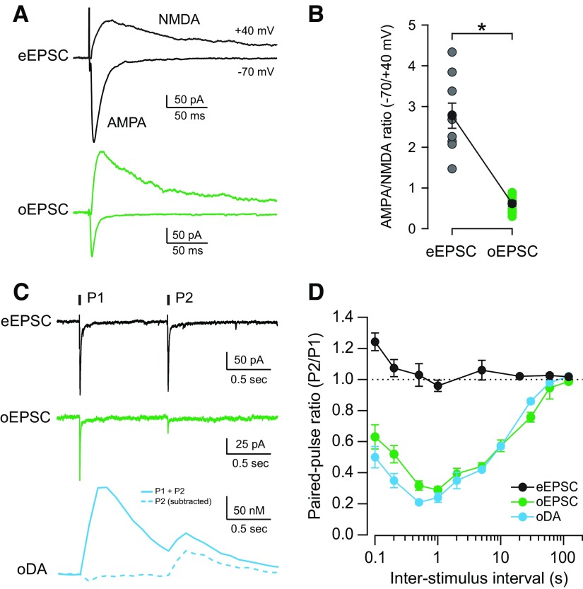
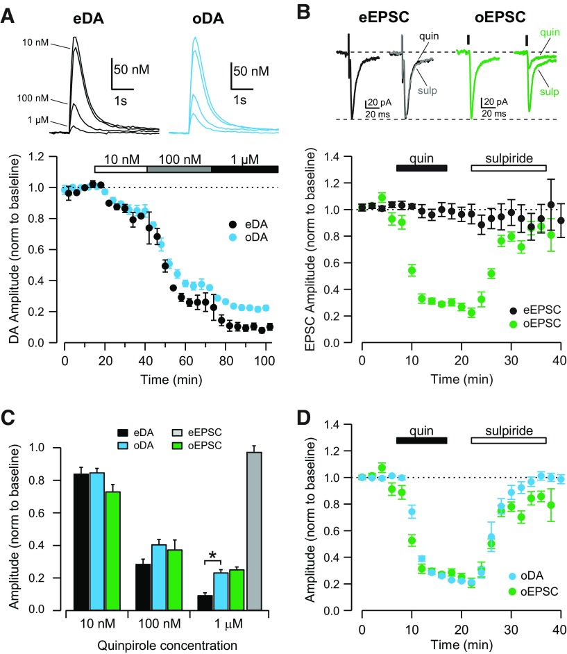
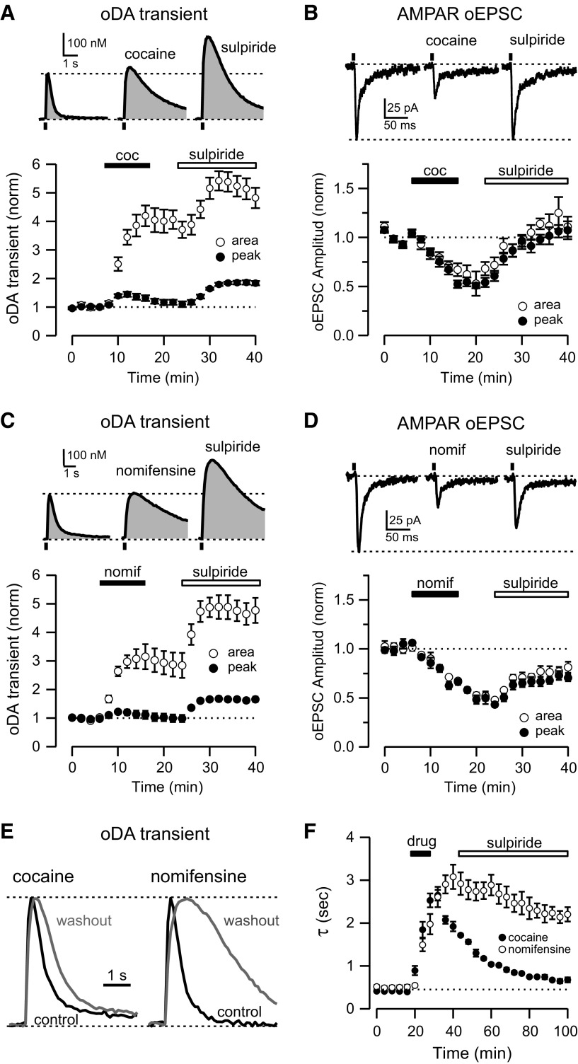
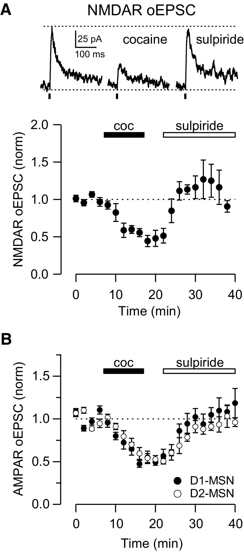
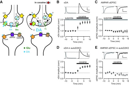
Synaptic transmission between ventral tegmental area and nucleus accumbens (NAc) is critically involved in reward-motivated behaviors and thought to be altered in addiction. In addition to dopamine (DA), glutamate is packaged and released by a subset of mesolimbic DA neurons, eliciting EPSCs onto medium spiny neurons in NAc. Little is known about the properties and modulation of glutamate release from DA midbrain terminals and the effect of cocaine. Using an optogenetic approach to selectively activate midbrain DA fibers, we compared the properties and modulation of DA transients and EPSCs measured using fast-scan cyclic voltammetry and whole-cell recordings in mouse brain slices. DA transients and EPSCs were inhibited by DA receptor D2R agonist and showed a marked paired-pulse depression that required 2 min for full recovery. Cocaine depressed EPSCs amplitude by 50% but enhanced the overall DA transmission from midbrain DA neurons. AMPA and NMDA receptor-mediated EPSCs were equally inhibited by cocaine, suggesting a presynaptic mechanism of action. Pharmacological blockage and genetic deletion of D2R in DA neurons prevented the cocaine-induced inhibition of EPSCs and caused a larger increase in DA transient peak, confirming the involvement of presynaptic D2R. These findings demonstrate that acute cocaine inhibits DA and glutamate release from midbrain DA neurons via presynaptic D2R but has differential overall effects on their transmissions in the NAc. We postulate that cocaine, by blocking DA reuptake, prolongs DA transients and facilitates the feedback inhibition of DA and glutamate release from these terminals.

Keywords: D2 receptors; co-release; cocaine; dopamine; feedback inhibition; glutamate.


Figures

Figure 1.
Figure 1.
DA transients evoked by electrical and optogenetic stimulation share common properties. A, Fluorescence image of sagittal brain section of DAT+/IRES–cre mouse showing labeled midbrain dopaminergic neurons expressing ChR2 and their projections to the NAc and dorsal striatum overlaid onto corresponding sections from the mouse brain atlas (Franklin and Paxinos, 2007). Scale bar, 1 mm. B, Confocal image of labeled cell bodies in the VTA. Scale bar, 50 μm. C, Confocal image in the NAc region showing dense innervation of labeled DA axons. No labeled cell bodies were found in the striatum. Scale bar, 50 μm. D, Top, Representative DA transients evoked by alternating electrical stimulation (eDA, black traces) and optogenetic stimulation (oDA, blue traces) recorded using FSCV in the NAc shell before and after TTX (500 nm) application. Bottom, Color plot voltammogram of eDA and oDA transients before TTX application. E, Superimposed examples of current–voltage plot of eDA and oDA signals showing the characteristic oxidation and reduction peaks of DA. Decay time constant (F; τ, mean ± SEM) and amplitude (G) of eDA and oDA transients obtained from the same slice preparation (n = 44 slices). AcbC, accumbens core; AcbSh, accumbens shell; CPu, caudate putamen; LV, lateral ventricle; SNC, substantia nigra pars compacta; SNR, substantia nigra pars reticulata; Tu, olfactory tubercle; VP, ventral pallidum.
Figure 2.
Figure 2.
Glutamatergic and dopaminergic responses evoked by optogenetic stimulation of midbrain DA terminals show similar release properties. A, Representative traces of eEPSCs (black) and oEPSCs (green) recorded from MSNs in the NAc shell. AMPAR-mediated currents were recorded when holding at −70 mV (downward) and NMDAR currents when holding at +40 mV (upward). B, AMPA/NMDA ratio of eEPSCs (gray) and oEPSCs (green) recorded from individual MSNs and the mean ± SEM of each group shown in black (n = 9, 10 cells; *p < 0.0001). C, Representative traces of AMPAR eEPSCs (black), AMPAR oEPSCs (green), and oDA transients (blue) evoked by paired-pulse stimulation with 1 s interval. oDA transient evoked by the second pulse (dotted) was calculated by subtracting the single-pulse oDA transients for intervals between 0.1 and 2 s. D, PPRs (P2/P1) for AMPAR eEPSCs (black), AMPAR oEPSCs (green), and oDA transients (blue) were plotted as a function of ISI (n = 7–13 cells and 6–45 slices).
Figure 3.
Figure 3.
D2-like receptor agonist inhibits dopaminergic and glutamatergic transmission from the VTA. A, Top, Representative traces of eDA (black) and oDA (blue) transients recorded before and after the application of quinpirole at 10 nm, 100 nm, and 1 μm. Bottom, Time course showing the inhibition of eDA (black) and oDA (blue) transients by increasing concentrations of quinpirole (n = 7 slices). B, Top, Representative traces of eEPSCs (black) and oEPSCs (green) recorded before and after application of quinpirole (quin; 1 μm) and subsequently sulpiride (sulp; 1 μm). Bottom, Time course of the quinpirole effect on eEPSCs (black) and oEPSCs (green) and subsequent sulpiride (1 μm) application (n = 9, 6 cells). C, Dose-dependent effect of quinpirole on the amplitude of eDA transients (black), oDA transients (blue), oEPSCs (green), and eEPSCs (gray; n = 7 slices, 9, 6 cells; *p = 0.000014, unpaired t test). D, Time course showing the inhibition of oDA transients (blue) and oEPSCs (green) by 1 μm quinpirole and subsequent reversal by 1 μm sulpiride application (n = 5 slices, 6 cells).
Figure 4.
Figure 4.
Cocaine enhances DA transients and inhibits glutamate transmission from VTA terminals. A, Top, Representative oDA transients recorded in control conditions (left), after 10 μm cocaine application (middle), and after subsequent 1 μm sulpiride application (right). Bottom, Time course of the change of oDA transient amplitude (solid) and area (open) during cocaine (coc) and subsequent sulpiride application (n = 6 slices). B, Top, Representative oEPSCs traces recorded in control conditions (left), after 10 μm cocaine application (middle), and after subsequent 1 μm sulpiride application (right). Bottom, Time course of the change of oEPSCs amplitude (solid) and area (open) during cocaine and subsequent sulpiride application (n = 19 cells). C, Top, Representative oDA transients recorded in control conditions (left), after 5 μm nomifensine application (middle),and after subsequent 1 μm sulpiride application (right). Bottom, Time course of the change of oDA transient amplitude (solid) and area (open) during nomifensine and subsequent sulpiride application (n = 5 slices). D, Top, Representative oEPSCs traces recorded in control conditions (left), after 5 μm nomifensine application (nomif; middle), and after subsequent 1 μm sulpiride application (right). Bottom, Time course of the change of oEPSCs amplitude (solid) and area (open) during nomifensine and subsequent sulpiride application (n = 5 cells). E, Representative oDA transients in control conditions (black) and 1 h after cocaine and nomifensine washout (gray). Peaks are normalized to highlight the change in the decay phase of the transients. F, Time course of the change in the decay time constant (τ) of oDA transients during drug (cocaine or nomifensine) application.
Figure 5.
Figure 5.
Cocaine inhibits NMDAR-mediated oEPSCs and also AMPAR-mediated oEPSCs in D1–MSNs and D2–MSNs. A, Top, Representative NMDAR-mediated oEPSCs in control conditions (left), after 10 μm cocaine application (middle), and after subsequent 1 μm sulpiride application (right). Bottom, Time course of the change of NMDAR-mediated oEPSCs amplitude during cocaine (coc) and subsequent sulpiride application recorded in MSNs in the NAc shell (n = 5 cells). B, Time course of the change of AMPAR-mediated oEPSCs amplitude during cocaine and subsequent sulpiride application recorded from D1–MSNs (solid) and D2–MSNs (open) in the NAc shell (n = 11, 8 cells).
Figure 6.
Figure 6.
Cocaine inhibits glutamate and DA release via activation of presynaptic D2Rs. A, Diagram showing a possible mechanism of action of cocaine on the synaptic terminals from the VTA that release dopamine (DA; blue square) and glutamate (Glu; green circle) or both. Under control conditions (left), DA and glutamate are released in response to action potentials and activate mainly postsynaptic receptors. DAT uptakes DA from the synaptic cleft, keeping extracellular DA concentration low. In the presence of cocaine (right), DAT is blocked and DA reuptake is inhibited (1), causing an increase in extracellular DA level (2), that leads to activation of presynaptic D2R (3) and subsequent inhibition of DA and glutamate release (4). Thus, as cocaine inhibits DA and Glu release via activation of presynaptic D2R, it causes opposite actions on the neurotransmission, increasing DA but inhibiting glutamate transmission. B, Top, Representative oDA transients recorded in sulpiride (left) and sulpiride + cocaine (right). Scale bars: 2 s, 100 nm. Bottom, Time course of the change of oDA transient amplitude during 10 μm cocaine application in the constant presence of 1 μm sulpiride (n = 7 slices). C, Top, Representative oEPSCs traces recorded in sulpiride (left) and sulpiride + cocaine (right). Bottom, Time course of the change of oEPSCs amplitude during 10 μm cocaine application in the constant presence of 1 μm sulpiride (n = 6 cells). Scale bars: 25 ms, 50 pA. D, Top, Representative oDA transients in autoDrd2KO mice recorded before (left) and after (right) application of cocaine. Scale bars: 2 s, 50 nm. Bottom, Time course of the change of oDA transient amplitude recorded in autoDrd2KO mice during 10 μm cocaine application (n = 7 slices). E, Top, Representative oEPSCs traces recorded in autoDrd2KO mice before (left) and after (right) cocaine. Scale bars: 25 ms, 25 pA. Bottom, Time course of the change of oEPSCs amplitude recorded in autoDrd2KO mice during 10 μm cocaine application (n = 7 cells). All data are mean ± SEM.

References

    1. Alsiö J, Nordenankar K, Arvidsson E, Birgner C, Mahmoudi S, Halbout B, Smith C, Fortin GM, Olson L, Descarries L, Trudeau LÉ, Kullander K, Lévesque D, Wallén-Mackenzie A. Enhanced sucrose and cocaine self-administration and cue-induced drug seeking after loss of VGLUT2 in midbrain dopamine neurons in mice. J Neurosci. 2011;31:12593–12603. doi: 10.1523/JNEUROSCI.2397-11.2011. - DOI - PMC - PubMed
    1. Bäckman CM, Malik N, Zhang Y, Shan L, Grinberg A, Hoffer BJ, Westphal H, Tomac AC. Characterization of a mouse strain expressing Cre recombinase from the 3′ untranslated region of the dopamine transporter locus. Genesis. 2006;44:383–390. doi: 10.1002/dvg.20228. - DOI - PubMed
    1. Bello EP, Mateo Y, Gelman DM, Noaín D, Shin JH, Low MJ, Alvarez VA, Lovinger DM, Rubinstein M. Cocaine supersensitivity and enhanced motivation for reward in mice lacking dopamine D2 autoreceptors. Nat Neurosci. 2011;14:1033–1038. doi: 10.1038/nn.2862. - DOI - PMC - PubMed
    1. Bérubé-Carrière N, Riad M, Dal Bo G, Lévesque D, Trudeau LE, Descarries L. The dual dopamine-glutamate phenotype of growing mesencephalic neurons regresses in mature rat brain. J Comp Neurol. 2009;517:873–891. doi: 10.1002/cne.22194. - DOI - PubMed
    1. Bérubé-Carrière N, Guay G, Fortin GM, Kullander K, Olson L, Wallén-Mackenzie Å, Trudeau LE, Descarries L. Ultrastructural characterization of the mesostriatal dopamine innervation in mice, including two mouse lines of conditional VGLUT2 knockout in dopamine neurons. Eur J Neurosci. 2012;35:527–538. doi: 10.1111/j.1460-9568.2012.07992.x. - DOI - PubMed

Publication types

MeSH terms