. 2014 Apr 18;289(16):11042-11058.
doi: 10.1074/jbc.M113.524421. Epub 2014 Feb 26.

A histidine aspartate ionic lock gates the iron passage in miniferritins from Mycobacterium smegmatis

Affiliations

A histidine aspartate ionic lock gates the iron passage in miniferritins from Mycobacterium smegmatis

Sunanda Margrett Williams et al. J Biol Chem. .

Abstract

Dps (DNA-binding protein from starved cells) are dodecameric assemblies belonging to the ferritin family that can bind DNA, carry out ferroxidation, and store iron in their shells. The ferritin-like trimeric pore harbors the channel for the entry and exit of iron. By representing the structure of Dps as a network we have identified a charge-driven interface formed by a histidine aspartate cluster at the pore interface unique to Mycobacterium smegmatis Dps protein, MsDps2. Site-directed mutagenesis was employed to generate mutants to disrupt the charged interactions. Kinetics of iron uptake/release of the wild type and mutants were compared. Crystal structures were solved at a resolution of 1.8-2.2 Å for the various mutants to compare structural alterations vis à vis the wild type protein. The substitutions at the pore interface resulted in alterations in the side chain conformations leading to an overall weakening of the interface network, especially in cases of substitutions that alter the charge at the pore interface. Contrary to earlier findings where conserved aspartate residues were found crucial for iron release, we propose here that in the case of MsDps2, it is the interplay of negative-positive potentials at the pore that enables proper functioning of the protein. In similar studies in ferritins, negative and positive patches near the iron exit pore were found to be important in iron uptake/release kinetics. The unique ionic cluster in MsDps2 makes it a suitable candidate to act as nano-delivery vehicle, as these gated pores can be manipulated to exhibit conformations allowing for slow or fast rates of iron release.

Keywords: DNA-binding Protein; Ion Channels; Ionic Cluster; Iron; Iron Oxidation; Mycobacterium; Mycobacterium smegmatis; X-ray Crystallography.


Figures
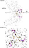
FIGURE 1.
FIGURE 1.
Ferritin-like pore harbors a funnel shaped channel leading into the protein cavity. A, the MsDps2 protein has a dodecameric form of which three subunits form the ferritin-like interface lined by many conserved negatively charged amino acids, thought to be crucial for iron uptake/release. The pathway for iron entry is represented by the arrows. The iron is further oxidized and stored as a ferric hydroxide mineral. B, the residues mutated in the current study are highlighted: magenta represents the amino acids at the widest region of the pore facing the solvent side; yellow shows the residues lining the narrowest point of the pore and opening into the protein cavity.
FIGURE 2.
FIGURE 2.
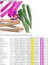
The histidine aspartate ionic lock in MsDps2 and structure-based sequence alignment with known Dps sequences. A, the 3-fold pore is shown with the residues comprising the ionic lock shown as spheres. The three subunits are shown in magenta, beige, and green; the corresponding residues are also colored in the same way. The nitrogen atoms in histidine 141 are blue, and the carboxyl group of aspartate 138 is shown in red. B, the residues at the outer surface are marked in yellow and the inner residues are in magenta. The histidines are not conserved in Dps homologs, but the aspartates at these positions are highly conserved (Asp138), and in the case of Asp127, substitutions are seen by glutamates in some cases.
FIGURE 3.
FIGURE 3.
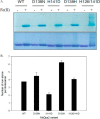
A, gel-based iron binding assay. MsDps2, D138N, H141D, D138H, and H126D/H141D proteins were loaded onto a 10% native PAGE, with (+) or without (−) added iron. The gel was stained with potassium ferricyanide (top panel), destained, and further stained with Coomassie Blue (bottom panel) to detect all the proteins. B, iron assimilated by the proteins in the expression and purification steps. After purification, the proteins were treated with sodium dithionite and 2,2′-bipyridyl, which forms a complex, [Fe(2,2′-bipyridyl)32+] with Fe(II). This complex can be assayed at 522 nm and the number of iron atoms assimilated per dodecamer was calculated and is given as a bar graph. Numerical values are given in the text. The values are averaged over 4–5 experiments with 2–3 different protein preparations.
FIGURE 4.
FIGURE 4.
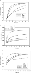
A, comparison of ferroxidation kinetics among the MsDps2 and mutants. Graphical representation of the progress curves showing ferroxidation for the various proteins as a function of time. Ferroxidation is triggered by the addition of ferrous sulfate to the protein solutions and monitored for a period of 60 min. The specific activity thus calculated is given in Table 3. B, comparison of iron release kinetics among MsDps2 and its variants. The rate of release of iron from proteins loaded with iron to half-capacity is monitored in a stopped flow instrument for a time period of 15 s to calculate the initial rate of release. The release was initiated by the addition of sodium dithionite (reduces Fe(III) to Fe(II)) and subsequent chelation by 2,2′-bipyridyl to form a pink colored complex with absorbance maxima at 522 nm. C, shows the reaction followed to 20 min where all iron is exited from the pores (except in case of D138H). This was used to calculate the time taken for the reduction and complexing of 60% of iron from the various proteins. The numerical values are shown in Table 3.
FIGURE 5.
FIGURE 5.
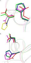
Superposition of the proteins with mutated residues on MsDps2 showing side chain conformational changes. The following color codes have been given for the residues from the various proteins; MsDps2, green; D138N, blue; H141D, magenta; D138H, yellow; and H126D/H141D, red. A, the residues of the mutant proteins has been overlaid with Asp138 and His141 from the MsDps2. The residues at position 141 remains more or less unaltered, with a translational shift exhibited by mutated aspartates in H141D and H126D/H141D. Mutations at the 138th position alter the side chain conformation in all cases. B, residues at 126 and 127 positions superposed on the MsDps2 His126 and Asp127 amino acids. Major shifts in side chain conformations are exhibited by mutants H141D and H126D/H141D at the 126th position, as described in the text.
FIGURE 6.
FIGURE 6.
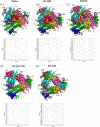
Prediction of interface clusters from the 3-fold interface with graph theory. A–E, interface clusters in the Protein Structure graphs (PSG6) of the ferritin-like interface of the MsDps2 (A, MsDps2 protein (PDB 2z90) and mutants; B, D138H (PDB 4M34); C, D138N (PDB 4M32); D, H126/141D (PDB 4M35); and E, H141D (PDB 4M33)) are represented as van der Waals spheres of each color representing a separate cluster. The pore clusters are absent in H141D and H126D/H141D variants. The residue composition of each cluster (vertical axis) for each cluster (horizontal axis) are provided below each of the schematic representations. Presence of a residue in a cluster is marked by a black dot. The clusters are named from C1 to C12 based on their occurrence in the respective PSG and are arbitrary.
FIGURE 7.
FIGURE 7.
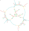
PSG of Ms-Dps2 ferritin-like interface (native and mutant). A comparison is shown of the native protein and mutant H126D/H141D showing the highest rate of iron release. Here the interactions of the ferritin-like trimeric interface are represented as nodes and edges. Each node represents an amino acid (naming nomenclature, [Chain][Residue number][Residue type]) and the edges are colored based on their interaction strength. The table below shows the color code for each type of node. This helps us in deeper understanding of the effects of mutations (B, H126D/H141D) on MsDps2 Ferritin-like interface (A, native). The residues mutated in H126D/H141D are assigned red font color. Only the interactions important in maintaining the pore interface are labeled for clarity. Asp130 is omitted in the mutant protein due to the absence of any interactions. Interaction strength (Equation 4): dark red, I > 8; red, 8 > I > 7; green, 7 > I > 6; yellow, 6 > I > 5; deep sky blue, 5 > I > 4; cyan, 4 > I > 3.
FIGURE 7.
FIGURE 7.
PSG of Ms-Dps2 ferritin-like interface (native and mutant). A comparison is shown of the native protein and mutant H126D/H141D showing the highest rate of iron release. Here the interactions of the ferritin-like trimeric interface are represented as nodes and edges. Each node represents an amino acid (naming nomenclature, [Chain][Residue number][Residue type]) and the edges are colored based on their interaction strength. The table below shows the color code for each type of node. This helps us in deeper understanding of the effects of mutations (B, H126D/H141D) on MsDps2 Ferritin-like interface (A, native). The residues mutated in H126D/H141D are assigned red font color. Only the interactions important in maintaining the pore interface are labeled for clarity. Asp130 is omitted in the mutant protein due to the absence of any interactions. Interaction strength (Equation 4): dark red, I > 8; red, 8 > I > 7; green, 7 > I > 6; yellow, 6 > I > 5; deep sky blue, 5 > I > 4; cyan, 4 > I > 3.
FIGURE 8.
FIGURE 8.
Van der Waals representation of the trimeric interface showing pore dimensions. A–E, the opening of the pore is measured by lines connecting the atoms nearest to each other in the pore to give approximate measurements of size in each case. A, MsDps2 protein (PDB 2Z90); B, D138N (PDB 4M32); C, H141D (PDB 4M33); D, D138H (PDB 4M34); and E, H126D/H141D (PDB 4M35). The gating residues are represented as sticks; the subunits of the trimer are colored blue, green, and magenta.
FIGURE 9.
FIGURE 9.
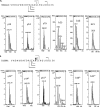
Expansion of CID MS/MS spectra of the precursor [M + 2H]2+ showing the difference of 1 Da between m/z values of b15, b16, b21, y13, y14, and y15 of MsDps2 (A) and D138N (B). The signals of b15, b16, b21, y13, y14, and y15 are shown with their isotopic peaks. The theoretical m/z values of MsDps2 are: b15 = 1536.6, b16 = 1649.7, b21 = 2185.91, y13 = 1393.74, y14 = 1495.82, y15 = 1582.97; D138N: b15 = 1535.69.6, b16 = 1648.78, b21 = 2184.91, y13 = 1392.79, y14 = 1493.78, and y15 = 1581.85. The ions having the mutated residue are indicated by an asterisk.
FIGURE 10.
FIGURE 10.
Model demonstrating the gating mechanism at the ferritin-like pore in MsDps2. Both figures are represented by their van der Waals radii. A shows the pore in its “closed” conformation. This is obtained from the MsDps2 PDB structure. B, the pore is in its open conformation. This was obtained by placing the MsDps2 gating residues in the side chain configurations exhibited by the H126D/H141D mutant, which showed the highest rate of iron release.

Similar articles

Cited by

References

    1. Aisen P., Enns C., Wessling-Resnick M. (2001) Chemistry and biology of eukaryotic iron metabolism. Int. J. Biochem. Cell Biol. 33, 940–959 - PubMed
    1. Benjamin J. A., Desnoyers G., Morissette A., Salvail H., Massé E. (2010) Dealing with oxidative stress and iron starvation in microorganisms: an overview. Can. J. Physiol. Pharmacol. 88, 264–272 - PubMed
    1. Bevers L. E., Theil E. C. (2011) Maxi- and mini-ferritins: minerals and protein nanocages. Prog. Mol. Subcell. Biol. 52, 29–47 - PMC - PubMed
    1. Bozzi M., Mignogna G., Stefanini S., Barra D., Longhi C., Valenti P., Chiancone E. (1997) A novel non-heme iron-binding ferritin related to the DNA-binding proteins of the Dps family in Listeria innocua. J. Biol. Chem. 272, 3259–3265 - PubMed
    1. Zhao G., Ceci P., Ilari A., Giangiacomo L., Laue T. M., Chiancone E., Chasteen N. D. (2002) Iron and hydrogen peroxide detoxification properties of DNA-binding protein from starved cells. A ferritin-like DNA-binding protein of Escherichia coli. J. Biol. Chem. 277, 27689–27696 - PubMed

Publication types

MeSH terms