. 2014 Apr 11;289(15):10551-10565.
doi: 10.1074/jbc.M113.538892. Epub 2014 Feb 25.

Insight into the roles of helicase motif Ia by characterizing Fanconi anemia group J protein (FANCJ) patient mutations

Affiliations

Insight into the roles of helicase motif Ia by characterizing Fanconi anemia group J protein (FANCJ) patient mutations

Manhong Guo et al. J Biol Chem. .

Abstract

Helicases are molecular motors that couple the energy of ATP hydrolysis to the unwinding and remodeling of structured DNA or RNA, which is coordinated by conserved helicase motifs. FANCJ is a DNA helicase that is genetically linked to Fanconi anemia, breast cancer, and ovarian cancer. Here, we characterized two Fanconi anemia patient mutations, R251C and Q255H, that are localized in helicase motif Ia. Our genetic complementation analysis revealed that both the R251C and Q255H alleles failed to rescue cisplatin sensitivity of a FANCJ null cell line as detected by cell survival or γ-H2AX foci formation. Furthermore, our biochemical assays demonstrated that both purified recombinant proteins abolished DNA helicase activity and failed to disrupt the DNA-protein complex. Intriguingly, R251C impaired DNA binding ability to single-strand DNA and double-strand DNA, whereas Q255H retained higher binding activity to these DNA substrates compared with wild-type FANCJ protein. Consequently, R251C abolished its DNA-dependent ATP hydrolysis activity, whereas Q255H retained normal ATPase activity. Physically, R251C had reduced ATP binding ability, whereas Q255H had normal ATP binding ability and could translocate on single-strand DNA. Although both proteins were recruited to damage sites in our laser-activated confocal assays, they lost their DNA repair function, which explains why they exerted a domain negative effect when expressed in a wild-type background. Taken together, our work not only reveals the structural function of helicase motif Ia but also provides the molecular pathology of FANCJ in related diseases.

Keywords: DNA Helicase; DNA Repair; Enzymes; FANCJ; Fanconi Anemia; Helicase; Motif Ia; Mutant; Pathogenesis.


Figures
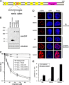
FIGURE 1.
FIGURE 1.
Expression of FANCJ-R251C or FANCJ-Q255H fails to rescue sensitivity of fancj mutant cells to the DNA cross-linker cisplatin. Panel A, schematic depicting FANCJ protein with the conserved helicase core domain and position of the FA causing mutations in motif Ia. The Fe-S domain and BRCA1 binding domain are indicated. Panel B, Western blot analysis of wild-type DT40 cells or fancj null cells transfected with plasmids encoding GFP, GFP-FANCJ-WT, GFP-FANCJ-R251C, or GFP-FANCJ-Q255H. Protein was detected with antibody against GFP (1:1000, Clontech) or actin (as a loading control, 10% loaded). Panel C, cisplatin sensitivity of cells with the indicated genotypes was evaluated by colony formation assay. Panel D, γ-H2AX immunofluorescence staining of fancj null cells transfected with plasmids encoding FANCJ-WT or FANCJ-R251C or FANCJ-Q255H. DT40 cells were treated with or without 1 μm cisplatin for 12 h followed by immunofluorescence detection as described under “Experimental Procedures.” Panel E, quantitative analyses of γ-H2AX foci in the corresponding transfected fancj null cell lines shown in panel D. Data represent the mean of at least 100 cells counted, with the S.D. indicated by error bars. Using a Student's t test for analysis of the γ-H2AX foci data (cisplatin treated), the differences in p values between fancj−/− FANCJ-WT cells and fancj−/− FANCJ-R251C or fancj−/− FANCJ-Q255H were 0.0002 and 0.00005, respectively, indicating a significant difference (p < 0.01) for each.
FIGURE 2.
FIGURE 2.
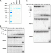
Helicase assays of FANCJ wild-type and mutants on forked duplex and G-quadruplex DNA substrates. Panel A, the purity of the wild-type and mutant FANCJ proteins was evaluated by their detected migration after SDS-PAGE on Coomassie-stained gels according to their predicted sizes. Two micrograms were loaded for each protein. Panel B, helicase reactions (20 μl) were performed by incubating the indicated FANCJ concentration with 0.5 nm forked duplex DNA substrate at 30 °C for 15 min as described under “Experimental Procedures.” Triangle, heat-denatured DNA substrate control. Panel C, helicase reactions were performed as for panel B, except G4 DNA was used instead of forked duplex. M, radiolabeled TP-G4 49-mer oligonucleotide (supplemental Table S1) marker.
FIGURE 3.
FIGURE 3.
R251C and Q255H mutants are compromised in their ability to displace DNA-bound protein. The indicated concentrations of FANCJ-WT or R251C or Q255H proteins were incubated with 2 mm ATP and DNA substrate (0.5 nm), which had streptavidin bound to the covalently linked biotin moiety residing 52 nucleotides from the 5′end of the radiolabeled oligonucleotide (supplemental Table S1).
FIGURE 4.
FIGURE 4.
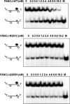
DNA binding of mutants and wild-type FANCJ proteins as detected by gel mobility shift assays. Panels A and B, the indicated concentrations of FANCJ-WT, -R251C, or -Q255H protein were incubated with 0.5 nm forked duplex DNA (panel A) or 44-mer ssDNA Tstem25 (panel B) substrate at room temperature for 30 min as described under “Experimental Procedures.” The DNA-protein complexes were resolved on native 5% polyacrylamide gels. Quantitative analyses of DNA gel-shift experiments are shown on the right. Panel C, 2.4 nm FANCJ-WT or -Q255H was incubated with 0.5 nm radiolabeled forked duplex DNA at room temperature for 15 min, and the indicated concentrations of 45-mer ssDNA (dT45) were subsequently added and incubated for an additional 15 min at room temperature. DNA-protein complexes were resolved on native 5% polyacrylamide gels. Quantitative analyses of DNA binding data from DNA competition experiments are shown on the right, with the S.D. indicated by error bars.
FIGURE 5.
FIGURE 5.
ATP binding by wild-type FANCJ and mutants proteins. [α-32P]ATP binding to FANCJ-WT and mutants was performed by gel filtration chromatography as described under “Experimental Procedures.” The same amount of protein was used, and the total amount of bound ATP was divided by protein and presented as mol ATP/pmol of protein. BSA was used as a control. Statistical analysis of the ATP binding data demonstrated that the difference in p value between FANCJ-WT and -R251C was 0.0007, indicating statistical significance (p < 0.01). The difference in p value between FANCJ-WT and -Q255H was 0.052, indicating that it was not statistically significant (p > 0.05).
FIGURE 6.
FIGURE 6.
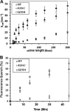
Translocase activity of wild-type FANCJ and mutants proteins. Panel A, the kcat values for ATP hydrolysis catalyzed by FANCJ proteins were determined as a function of oligonucleotide dT length, as described under “Experimental Procedures.” The oligonucleotide dT (length 5–200 nucleotides) concentration ranged from 50 to 5000 nm depending on the length of the oligo dT tested. Error bars represent the S.D. of the fit to the Michaelis-Menten equation. Panel B, fluorescence quenching experiments, performed as described under “Fluorescence Quench Translocation Assays,” using FANCJ-WT and mutants.
FIGURE 7.
FIGURE 7.
Size exclusion chromatography and biochemical analysis of selected FANCJ protein fractions. Panel A, chromatographic profiles of recombinant FANCJ-WT, -R251C, and -Q255H eluting from a Superdex200 HR gel filtration column. Panel B, helicase reactions (20 μl) were performed by incubating the indicated FANCJ fractions with 0.5 nm DNA substrate at 30 °C for 15 min as described under “Experimental Procedures.” Triangle, heat-denatured DNA substrate control. Panel C, ATP hydrolysis (kcat) was determined for each fraction in the presence of covalently closed M13 single-strand DNA (2.1 nm). Statistical analysis of the ATPase data for the B9/10 fraction demonstrated that the difference in p value between FANCJ-WT and -R251C was 0.0001, indicating statistical significance (p < 0.01). The difference in p value between FANCJ-WT and -Q255H was 0.313, indicating that it was not statistically significant (p > 0.05). Similar results were obtained for the three B6 fractions. Panel D, indicated concentrations of FANCJ proteins were incubated with 0.5 nm forked duplex DNA substrate at room temperature for 30 min under standard gel shift assay conditions as described under “Experimental Procedures.” DNA-protein complexes were resolved on native 5% polyacrylamide gels.
FIGURE 8.
FIGURE 8.
DNA damage response of wild-type and mutants FANCJ proteins in cells. Panel A, GFP fluorescence detection was performed on HeLa cells transfected with plasmids encoding GFP-FANCJ-WT, GFP-FANCJ-R251C, or GFP-FANCJ-Q255H. Cells were treated with 10 mm hydroxyurea (HU) or 1 μm mitomycin C (MMC) for 12 h and fixed for observation under microscope. Panel B, quantitative analyses of GFP-FANCJ foci formation. Data represent the mean of at least 100 cells counted, with the S.D. indicated by error bars. The difference in p value between WT and R251C or Q255H was not statistically significant (p > 0.05) under the three conditions. Panel C, GFP-FANCJ-transfected HeLa cells were treated with a laser (5.5%) at defined regions to induce localized DSBs or incubated with psoralen followed by laser treatment (1.8%) to induce Pso-ICLs. GFP-FANCJ recruitment to Pso-ICLs or DSBs was observed by live-cell imaging using confocal immunofluorescence microscopy 5 min after laser-induced damage.
FIGURE 9.
FIGURE 9.
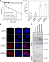
Expression of FANCJ-R251C or FANCJ-Q255H mutant exerts a dominant negative effect on wild-type cells. Panel A, cisplatin sensitivity of cells with indicated genotypes was evaluated by colony formation assay. Panel B, γ-H2AX immunofluorescence staining of cisplatin-treated fancj+/+ cells transfected with plasmids encoding GFP, GFP-FANCJ-WT, GFP-FANCJ-R251C, or GFP-FANCJ-Q255H. DT40 cells were treated with 1 μm cisplatin for 12 h followed by immunofluorescence detection. Panel C, quantitative analyses of γ-H2AX foci shown in Panel B. Data represent the mean of at least 100 cells counted, with the S.D. indicated by error bars. Statistical analysis of the γ-H2AX foci data demonstrated that the differences in p values between fancj+/+ FANCJ-WT cells and fancj+/+ FANCJ-R251C or fancj+/+ FANCJ-Q255H were 0.004 and 0.005, respectively, indicating a significant difference (p < 0.01) for each. Panel D, co-immunoprecipitation experiments using nuclear extracts from HeLa cells expressing GFP, GFP-FANCJ-WT, GFP-FANCJ-R251C, or GFP-FANCJ-Q255H and immunoprecipitated with GFP antibody (GFP-IPed) with the pulldown detected with antibodies against TopBP1, MLH1, and BRCA1, respectively.
FIGURE 10.
FIGURE 10.
Alignment of helicase motif Ia and model of human FANCJ structure. Panel A, alignment of helicase motif Ia from helicases NPH-II, Prp18p, Prp22, and Prp43. Panel B, mutation sites in the modeled FANCJ structure that are based on SaXPD (23). The helicase domains HD1 (cyan) and HD2 (green), the Fe-S domain (orange), and the Arch domain (purple) are indicated. The position of the pore formed by the four domains is noted. The mutated residues are marked in red and indicated with red arrows. The black line represents DNA, including double helix and ssDNA, and the dashed lines represent DNA behind the protein. Enlargement of the local structure harboring the Arg-251 and Gln-255 residues is shown on the right. Panel C, alignment of helicase motif Ia for FANCJ-sequence like helicases.

Similar articles

Cited by

References

    1. Bernstein K. A., Gangloff S., Rothstein R. (2010) The RecQ DNA helicases in DNA repair. Annu. Rev Genet. 44, 393–417 - PMC - PubMed
    1. Dillingham M. S. (2011) Superfamily I helicases as modular components of DNA-processing machines. Biochem. Soc. Trans. 39, 413–423 - PubMed
    1. Lohman T. M., Bjornson K. P. (1996) Mechanisms of helicase-catalyzed DNA unwinding. Annu. Rev. Biochem. 65, 169–214 - PubMed
    1. Lohman T. M., Tomko E. J., Wu C. G. (2008) Non-hexameric DNA helicases and translocases. Mechanisms and regulation. Nat. Rev. Mol. Cell Biol. 9, 391–401 - PubMed
    1. Fairman-Williams M. E., Guenther U. P., Jankowsky E. (2010) SF1 and SF2 helicases. Family matters. Curr. Opin. Struct. Biol. 20, 313–324 - PMC - PubMed

Publication types

MeSH terms