. 2014 Apr 18;289(16):11132-11142.
doi: 10.1074/jbc.M113.492512. Epub 2014 Feb 26.

Antibodies that detect O-linked β-D-N-acetylglucosamine on the extracellular domain of cell surface glycoproteins

Affiliations

Antibodies that detect O-linked β-D-N-acetylglucosamine on the extracellular domain of cell surface glycoproteins

Yuko Tashima et al. J Biol Chem. .

Abstract

The transfer of N-acetylglucosamine (GlcNAc) to Ser or Thr in cytoplasmic and nuclear proteins is a well known post-translational modification that is catalyzed by the O-GlcNAc transferase OGT. A more recently identified O-GlcNAc transferase, EOGT, functions in the secretory pathway and transfers O-GlcNAc to proteins with epidermal growth factor-like (EGF) repeats. A number of antibodies that detect O-GlcNAc in cytosolic and nuclear extracts have been described previously. Here we compare seven of these antibodies (CTD110.6, 10D8, RL2, HGAC85, 18B10.C7(#3), 9D1.E4(#10), and 1F5.D6 (#14) for detection of the O-GlcNAc modification on extracellular domains of membrane or secreted glycoproteins that may also carry various N- and O-glycans. We found that CTD110.6 binds not only to O-GlcNAc on proteins but also to terminal β-GlcNAc on the complex N-glycans of Lec8 Chinese hamster ovary (CHO) cells that lack UDP-Gal transporter activity and express GlcNAc-terminating, complex N-glycans. We show that CTD110.6, #3, and #10 antibodies can be used to detect cell surface glycoproteins bearing O-GlcNAc. Cell surface glycoproteins recognized by CTD110.6 antibody included NOTCH1 that possesses many EGF repeats with a consensus site for EOGT. Knockdown of CHO Eogt reduced binding of CTD110.6 to Lec1 CHO cells, and expression of a human EOGT cDNA increased the O-GlcNAc signal on Lec1 cells and the extracellular domain of NOTCH1. Thus, with careful controls, antibodies CTD110.6 (IgM), #3 (IgG), and #10 (IgG) can be used to detect membrane and secreted proteins modified by O-GlcNAc on EGF repeats.

Keywords: Antibodies; CHO; Flow Cytometry; GlcNAc-terminating N-Glycans; Lec1; Lec8; Mutant; O-GlcNAc; O-GlcNAcylation.


Figures

FIGURE 1.
FIGURE 1.
Antibody CTD110.6 binds to terminal β-GlcNAc in complex N-glycans. A, Western blot (IB) analysis of LFNG-AP and MFNG-AP affinity-purified using anti-PLAP antibody-conjugated agarose from conditioned medium of Lec2/vector (Lec2/V), Lec2/Lfng-AP (Lec2/L), Lec2/Mfng-AP (Lec2/M), Lec8/vector (Lec8/V), Lec8/Lfng-AP (Lec8/L), or Lec8/Mfng-AP (Lec8/M) CHO cells. After separation on a 7.5% SDS-polyacrylamide gel and transfer, PVDF membranes were incubated at room temperature for 1 h with mAbs CTD110.6 (0.4 μg/ml) and anti-mouse IgM antibody-HRP (1.6 μg/ml), or anti-human PLAP (L-19) antibody (1 μg/ml) and anti-goat IgG antibody-HRP (0.08 μg/ml). Representative results are of blots from three independent experiments. B, Lec8/V and Lec8/M cells transiently transfected with pMirb/Mfng-AP. MFNG-AP affinity-purified using anti-FLAG (M2) antibody and protein G beads from conditioned medium were analyzed on a 7.5% SDS-polyacrylamide gel and detected by Western blot analysis as in A. U, Unbound; B, Bound. Representative results are of blots from three independent experiments. C, MFNG-HA-FLAG collected from conditioned medium of Lec8/V and Lec8/M cells transiently expressing pcDNA3/Mfng-HA-FLAG using anti-FLAG (M2) beads, separated in a 7.5% SDS-polyacrylamide gel, and subjected to Western blot analysis with CTD110.6 as in A or anti-FLAG (M2) antibody (1 μg/ml) and anti-mouse IgG antibody-HRP (0.08 μg/ml). Representative results are of blots from three independent experiments. D, MFNG-AP collected from conditioned medium of Lec8/M and Lec2/M cells treated with PNGase F or β-N-acetylhexosaminidase followed by Western blot analysis. E, Lec8/V and Lec8/M cells cultured with or without 5 μg/ml swainsonine for 4 days. MFNG-AP collected from conditioned media was analyzed by SDS-PAGE and Western blot analysis as in A, before and after treatment with PNGase F.
FIGURE 2.
FIGURE 2.
Detection of GlcNAc-terminated, complex N-glycans by other anti-O-GlcNAc antibodies. A, MFNG-AP affinity-purified from Lec8/M conditioned medium (upper) or Lec8/M whole cell lysate (lower) was separated in a 10% SDS-polyacrylamide gel, transferred to PVDF membranes, and incubated with anti-O-GlcNAc antibodies (top and bottom), or anti-human PLAP (L-19) antibody (middle), for 1 h at room temperature. O-GlcNAcylated proteins were detected with CTD110.6 (0.4 μg/ml) or 10D8 (0.2 μg/ml) and anti-mouse IgM antibody-HRP (1.6 μg/ml), RL2 (3 μg/ml), HGAC85 (4 μg/ml), #3 (0.86 μg/ml), #10 (0.59 μg/ml), or #14 (0.97 μg/ml) and anti-mouse IgG-HRP (0.08 μg/ml). Representative results are from two preparations. B, mAbs #3 (8.6 μg/ml) and #14 (9.7 μg/ml) were incubated at room temperature for 1 h followed by anti-mouse IgG-HRP (0.08 μg/ml). IP, immunoprecipitate; IB, immunoblot.
FIGURE 3.
FIGURE 3.
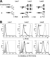
CTD110.6 recognizes both O-GlcNAc and terminal β-GlcNAc on cell surface glycoproteins. A, N-glycans of glycoproteins synthesized in CHO glycosylation mutants cultured in the presence (predicted (28, 29)), or absence (26, 27), of swainsonine. B, binding of L-PHA (upper) and mAb CTD110.6 (lower) to fixed cells. Lec2 and Lec8 cells were cultured with or without 5 μg/ml swainsonine (SW) for 4 days. Cells (4 × 10⁵) were incubated with 20 μg/ml fluorescein-labeled L-PHA or 40 μg/ml CTD110.6 on ice for 20 min followed by 10 μg/ml Cy5-conjugated anti-mouse IgM antibody. Gray profiles, secondary Abs; thin line, control cells; bold line, swainsonine-treated cells. Representative results are from two independent experiments.
FIGURE 4.
FIGURE 4.
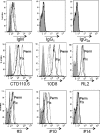
Binding of anti-O-GlcNAc antibodies to Lec1 cells. Lec1 cells (4 × 10⁵) fixed with 4% paraformaldehyde (Fix) or fixed and permeabilized with 0.1% Triton X-100 (Perm) were incubated with 10 μg/ml 10D8 or 40 μg/ml other anti-O-GlcNAc antibodies including isotype control Abs followed by incubation with 10 μg/ml secondary Ab. Gray profiles, secondary Ab; short dashed line, isotype control for Fix; long dashed line, isotype control for Perm; bold solid line, anti-O-GlcNAc mAb for Fix; thin solid line, anti-O-GlcNAc mAb for Perm. Representative results are from two independent experiments.
FIGURE 5.
FIGURE 5.
GlcNAc competes for cell surface binding of only some anti-O-GlcNAc mAbs. Lec1 cells (4 × 10⁵) fixed with 4% paraformaldehyde were incubated with 10 μg/ml 10D8 mAb or 40 μg/ml each of the other anti-O-GlcNAc mAbs (CTD110.6, RL2, #3, #10, and #14) in the presence of 5 mm GlcNAc (bold line) or 5 mm GalNAc (dotted line), or no sugar (thin line) on ice for 20 min followed by 10 μg/ml secondary Ab. Gray profiles, secondary Ab alone. Representative results are from four independent experiments.
FIGURE 6.
FIGURE 6.
Specificity of CTD110.6 for O-GlcNAc on cell surface glycoproteins. A, Lec1 cells were biotinylated, separated into SA-Bound and Unbound fractions on SA-agarose, and analyzed before and after treatment with PNGase F by SDS-PAGE and Western blotting (IB) using the CTD110.6 mAb. B, flow cytometry profile shows CTD110.6 mAb of fixed Lec1 cells expressing vector (thin line) or siRNA 1239 (bold line). Dashed line is isotype control. Representative results are from two independent experiments. C, results are same as B except the bold line is for Lec1 cells treated with a mixture of siRNA 1239 and 1233 (1:1). Representative results are from two independent experiments. D, Lec1 cells expressing vector control (thin line) or a human EOGT cDNA (bold line) were fixed and analyzed by flow cytometry with CTD110.6 mAb. Dashed line is isotype control. Representative results are from three independent experiments. E, Lec1 cells expressing vector control or human EOGT cDNA were analyzed by Western blotting using anti-EOGT antibody (1:500 at 4 °C overnight). Representative results are from two independent experiments.
FIGURE 7.
FIGURE 7.
O-GlcNAc on NOTCH1 is detected at the cell surface. A, Lec1 cells transiently expressing NOTCH1-Myc were treated for 18 h with 100 μm PUGNAc (+) or vehicle (dimethyl sulfoxide) (−), biotinylated, lysed, and the lysate was incubated with anti-Myc beads (Myc-Bound) or anti-IgG beads. The fraction that did not bind to anti-Myc or anti-IgG beads was incubated with SA-agarose and gave the (SA-Bound after first immunoprecipitation (IP)) and (SA-Unbound after immunoprecipitate-Myc) fractions. Samples were separated on a 7.5% SDS-polyacrylamide gel, and one set was probed with anti-NOTCH1 ECD mAb (2 μg/ml), stripped, and probed with anti-Myc mAb (1:500). The second set was probed with CTD110.6 (0.4 μg/ml). The stars identify proteins that migrated similarly to but distinct from NOTCH1-Myc. Arrowhead identifies band indicating equal loading. Representative results are from three independent experiments. IB, immunoblot. B, NOTCH1-Myc prepared from transient CHO transfectants as in A was incubated with 10 units of β-N-acetylhexosaminidase at 37 °C for 2 h, separated in a 7.5% SDS-polyacrylamide gel, transferred to PVDF membranes, and incubated with CTD110.6 (0.4 μg/ml) and anti-mouse IgM-HRP (1.6 μg/ml) or anti-Myc 9E10 (1:500) and anti-mouse IgG-HRP (0.08 μg/ml) Abs at room temperature for 1 h. Representative results are from two independent experiments. C, Lec1 cells were co-transfected with vector or pCR3.1/EOGT, and pCS2+/Notch1-Myc. After 24 h, biotinylation was performed; biotinylated proteins were collected on SA-agarose beads or by incubation with anti-Myc antibody and protein G beads, and analyzed by SDS-PAGE and Western blotting with CTD110.6 mAb as in A. D, lysate from Lec1 cells transfected with plasmid pCS2+ (V) or pCS2+/Notch1-Myc (N1) was precleared with anti-IgM-agarose, incubated with mAb CTD110.6 (1 μg) overnight at 4 °C, and collected on mouse anti-IgM-agarose. Lysate (50 μl) and proteins solubilized from beads were separated on a 7.5% SDS-polyacrylamide gel, transferred to membrane, and subjected to Western blot analysis using the indicated antibodies sequentially, in the order CTD110.6, anti-NOTCH1, and anti-Myc antibodies. Representative results are from three experiments.
FIGURE 8.
FIGURE 8.
O-GlcNAc is present on a range of CHO cell surface proteins. A, Lec1 cells were biotinylated, lysed, and biotinylated proteins were collected on SA-agarose. SA-Bound and Unbound proteins were separated in a 10% SDS-polyacrylamide gel. The PVDF membranes were incubated with anti-PDGFR-α (2 μg/ml) and anti-rabbit IgG-HRP (1:5000), anti-protein disulfide isomerase (2 μg/ml) or anti-β-actin (1:5000) Abs and anti-mouse IgG-HRP (0.16 μg/ml), or NeutrAvidin-HRP (1:5000) at room temperature for 1 h. Representative results are from four independent experiments. IB, immunoblot. B, samples were the same as in A, but run on a 7.5% SDS-polyacrylamide gel. Immunoblotting was with CTD110.6 (0.4 μg/ml) and anti-mouse IgM antibody-HRP (1.6 μg/ml), or anti-NOTCH3 (5E1) (1:500) and anti-mouse IgG-HRP (0.08 μg/ml) antibodies at room temperature for 1 h. Arrowheads identify O-GlcNAcylated cell surface glycoproteins. C, samples were the same as in A and run on a 10% SDS-polyacrylamide gel. Immunoblotting was with CTD110.6 (0.4 μg/ml) or #10 (0.59 μg/ml) antibodies at room temperature for 1 h. Arrowheads indicate bands present in the biotinylated, SA-Bound fraction. Representative results are from two independent experiments.

Similar articles

Cited by

References

    1. Varki A., Cummings R. D., Esko J. D., Freeze H. H., Stanley P., Bertozzi C. R., Hart G. W., Etlzler M. E. (2009) Essentials of Glycobiology, 2nd Edition, Cold Spring Harbor Laboratory, Cold Spring Harbor, NY - PubMed
    1. Matsuura A., Ito M., Sakaidani Y., Kondo T., Murakami K., Furukawa K., Nadano D., Matsuda T., Okajima T. (2008) O-Linked N-acetylglucosamine is present on the extracellular domain of Notch receptors. J. Biol. Chem. 283, 35486–35495 - PubMed
    1. Sakaidani Y., Furukawa K., Okajima T. (2010) O-GlcNAc modification of the extracellular domain of Notch receptors. Methods Enzymol. 480, 355–373 - PubMed
    1. Sakaidani Y., Ichiyanagi N., Saito C., Nomura T., Ito M., Nishio Y., Nadano D., Matsuda T., Furukawa K., Okajima T. (2012) O-Linked N-acetylglucosamine modification of mammalian Notch receptors by an atypical O-GlcNAc transferase Eogt1. Biochem. Biophys. Res. Commun. 419, 14–19 - PubMed
    1. Sakaidani Y., Nomura T., Matsuura A., Ito M., Suzuki E., Murakami K., Nadano D., Matsuda T., Furukawa K., Okajima T. (2011) O-Linked-N-acetylglucosamine on extracellular protein domains mediates epithelial cell-matrix interactions. Nat. Commun. 2, 583. - PubMed

Publication types

MeSH terms

Substances