

Three-dimensional paper-based model for cardiac ischemia
- PMID: 24574054
- PMCID: PMC4107065
- DOI: 10.1002/adhm.201300575
Three-dimensional paper-based model for cardiac ischemia
Abstract
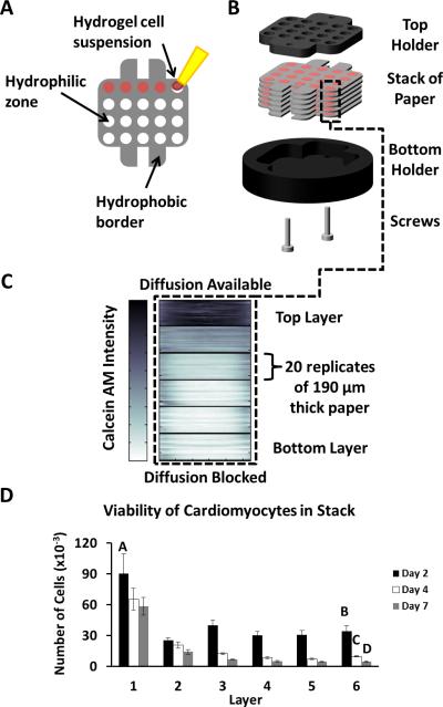
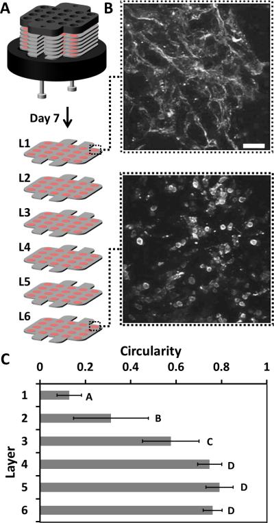
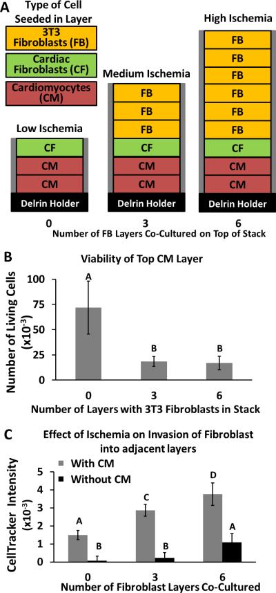
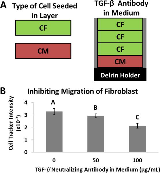
In vitro models of ischemia have not historically recapitulated the cellular interactions and gradients of molecules that occur in a 3D tissue. This work demonstrates a paper-based 3D culture system that mimics some of the interactions that occur among populations of cells in the heart during ischemia. Multiple layers of paper containing cells, suspended in hydrogels, are stacked to form a layered 3D model of a tissue. Mass transport of oxygen and glucose into this 3D system can be modulated to induce an ischemic environment in the bottom layers of the stack. This ischemic stress induces cardiomyocytes at the bottom of the stack to secrete chemokines which subsequently trigger fibroblasts residing in adjacent layers to migrate toward the ischemic region. This work demonstrates the usefulness of patterned, stacked paper for performing in vitro mechanistic studies of cellular motility and viability within a model of the laminar ventricle tissue of the heart.
Keywords: 3D cell culture; cardiac ischemia; cardiomyocytes; co-culture; gradients.
© 2014 WILEY-VCH Verlag GmbH & Co. KGaA, Weinheim.
Figures
Similar articles
-
Functional 3-D cardiac co-culture model using bioactive chitosan nanofiber scaffolds.Biotechnol Bioeng. 2013 Feb;110(2):637-47. doi: 10.1002/bit.24727. Epub 2012 Oct 5. Biotechnol Bioeng. 2013. PMID: 22991229
-
Age-dependent functional crosstalk between cardiac fibroblasts and cardiomyocytes in a 3D engineered cardiac tissue.Acta Biomater. 2017 Jun;55:120-130. doi: 10.1016/j.actbio.2017.04.027. Epub 2017 Apr 25. Acta Biomater. 2017. PMID: 28455218 Free PMC article.
-
Vascularized cardiac tissue construction with orientation by layer-by-layer method and 3D printer.Sci Rep. 2020 Mar 26;10(1):5484. doi: 10.1038/s41598-020-59371-y. Sci Rep. 2020. PMID: 32218447 Free PMC article.
-
Modeling Cardiovascular Diseases with hiPSC-Derived Cardiomyocytes in 2D and 3D Cultures.Int J Mol Sci. 2020 May 11;21(9):3404. doi: 10.3390/ijms21093404. Int J Mol Sci. 2020. PMID: 32403456 Free PMC article. Review.
-
Biophysical stimulation for in vitro engineering of functional cardiac tissues.Clin Sci (Lond). 2017 Jul 1;131(13):1393-1404. doi: 10.1042/CS20170055. Clin Sci (Lond). 2017. PMID: 28645929 Review.
Cited by
-
Myocardial infarction from a tissue engineering and regenerative medicine point of view: A comprehensive review on models and treatments.Biophys Rev (Melville). 2022 Sep;3(3):031305. doi: 10.1063/5.0093399. Epub 2022 Aug 30. Biophys Rev (Melville). 2022. PMID: 36091931 Free PMC article. Review.
-
Optically Active, Paper-Based Scaffolds for 3D Cardiac Pacing.ACS Appl Mater Interfaces. 2024 Oct 9;16(40):53449-53459. doi: 10.1021/acsami.4c10183. Epub 2024 Sep 27. ACS Appl Mater Interfaces. 2024. PMID: 39332816 Free PMC article.
-
Hydrogel-laden paper scaffold system for origami-based tissue engineering.Proc Natl Acad Sci U S A. 2015 Dec 15;112(50):15426-31. doi: 10.1073/pnas.1504745112. Epub 2015 Nov 30. Proc Natl Acad Sci U S A. 2015. PMID: 26621717 Free PMC article.
-
Mapping signalling perturbations in myocardial fibrosis via the integrative phosphoproteomic profiling of tissue from diverse sources.Nat Biomed Eng. 2020 Sep;4(9):889-900. doi: 10.1038/s41551-020-0585-y. Epub 2020 Jul 13. Nat Biomed Eng. 2020. PMID: 32661320
-
Increasing the packing density of assays in paper-based microfluidic devices.Biomicrofluidics. 2021 Feb 4;15(1):011502. doi: 10.1063/5.0042816. eCollection 2021 Jan. Biomicrofluidics. 2021. PMID: 33569089 Free PMC article. Review.
References
-
- Beanes SR, Dang C, Soo C, Ting K. Expert. Rev. Mol. Med. 2003;5:1. - PubMed
-
- Kuznetsov AV, Hermann M, Saks V, Hengster P, Margreiter R. Intern. J. Biochem. Cell Bio. 2009;41:1928. - PubMed
-
- Radisic M, Malda J, Epping E, Geng WL, Langer R, Vunjak-Novakovic G. Biotechnol. Bioeng. 2006;93:332. - PubMed
-
- Vanden Hoek TL, Shao Z, Li C, Zak R, Schumacker PT, Becker LB. Am. J. Physiol. 1996;270:H1334. - PubMed
Publication types
MeSH terms
Grants and funding
LinkOut - more resources
Full Text Sources
Other Literature Sources
