

Damage-associated molecular patterns stimulate interleukin-33 expression in nasal polyp epithelial cells
- PMID: 24574111
- PMCID: PMC3981922
- DOI: 10.1002/alr.21237
Damage-associated molecular patterns stimulate interleukin-33 expression in nasal polyp epithelial cells
Abstract
Background: Chronic rhinosinusitis with nasal polyps (CRSwNP) is a disorder characterized by eosinophilic inflammation and local T-helper 2 (Th2) cytokine production. Innate lymphoid cells that elaborate Th2 cytokines have recently been characterized within nasal polyps. These cells can be activated by the epithelial cell-derived cytokine interleukin-33 (IL-33). The objective of this study is to determine whether 2 molecules associated with tissue damage (high mobility group box-1 [HMGB-1] and adenosine triphosphate [ATP]) elicit expression of IL-33 in sinonasal epithelial cells (SNECs) derived from recalcitrant CRSwNP patients.
Methods: Ethmoid tissue was obtained from 8 recalcitrant CRSwNP and 9 control subjects during endoscopic sinus surgery (ESS). Tissue was prepared for immunohistochemistry and for SNEC air-liquid interface culture. After exposure to either HMGB1 or ATP in vitro, SNECs were processed for messenger RNA (mRNA) extraction and immunocytochemistry. IL-33 levels were determined by real-time polymerase chain reaction (PCR) and by immunochemical staining with anti-IL-33 antibody.
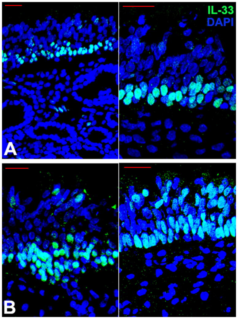
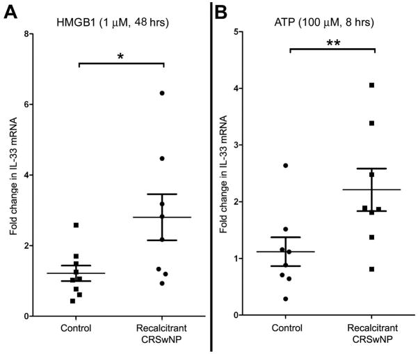
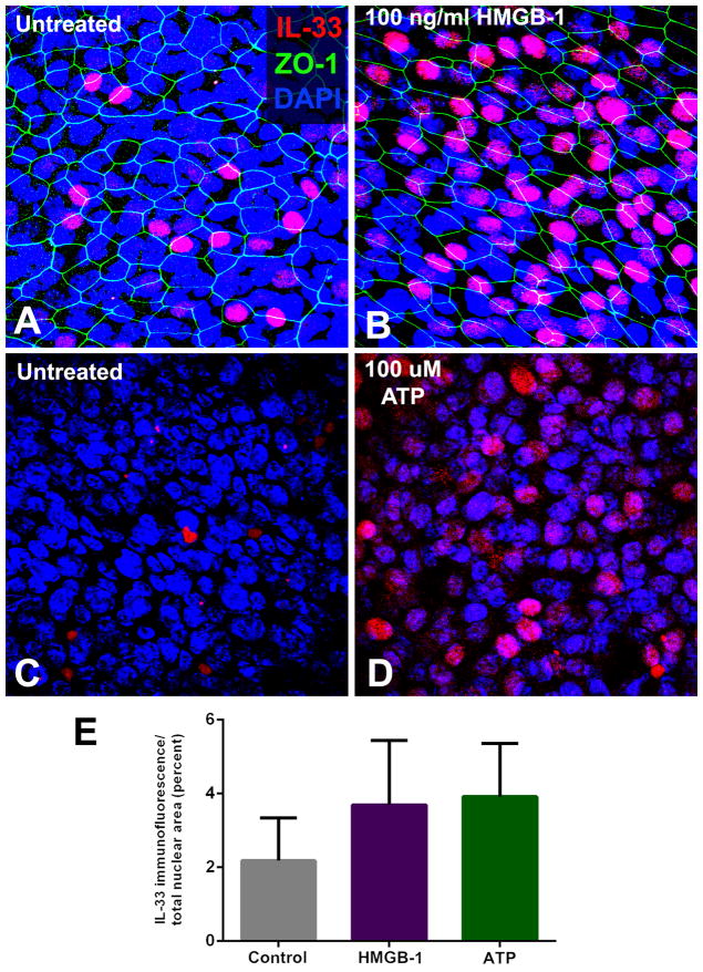
Results: Intranuclear IL-33 is normally expressed in basal epithelial cells, but is present in more apical cells and outside the nucleus in CRSwNP. Exposure of SNECs to HMGB-1 or ATP resulted in a statistically significant increase in IL-33 mRNA expression in SNECs derived from recalcitrant CRSwNP patients. This increase was reflected at the protein level by immunochemical staining of IL-33.
Conclusion: Tissue damage is a nonspecific trigger of epithelial IL-33 production in treatment-recalcitrant polyps, which may be responsible for perpetuating eosinophilic inflammation in CRSwNP. This common pathway may help explain why multiple environmental and infectious agents have been implicated in CRSwNP exacerbation.
Conflict of interest statement
Conflict-of-interest disclosure: The authors declare no competing financial interests.
Figures
References
-
- Fokkens WJ, Lund VJ, Mullol J, et al. EPOS 2012: European position paper on rhinosinusitis and nasal polyps 2012. A summary for otorhinolaryngologists. Rhinology. 2012 Mar;50(1):1–12. - PubMed
-
- Van Crombruggen K, Zhang N, Gevaert P, Tomassen P, Bachert C. Pathogenesis of chronic rhinosinusitis: Inflammation. J Allergy Clin Immunol. 2011 Oct;128(4):728–732. - PubMed
-
- Palmer JN. Bacterial biofilms: do they play a role in chronic sinusitis? Otolaryngol Clin North Am. 2005 Dec;38(6):1193–1201. viii. - PubMed
-
- Orlandi RR, Marple BF. Fungus and chronic rhinosinusitis: weighing the evidence. Otolaryngol Head Neck Surg. 2010 Nov;143(5):611–613. - PubMed
-
- Bachert C, van Zele T, Gevaert P, De Schrijver L, Van Cauwenberge P. Superantigens and nasal polyps. Curr Allergy Asthma Rep. 2003 Nov;3(6):523–531. - PubMed
Publication types
MeSH terms
Substances
Grants and funding
LinkOut - more resources
Full Text Sources
Other Literature Sources
Medical
