. 2014 May;88(9):5152-64.
doi: 10.1128/JVI.03851-13. Epub 2014 Feb 26.

Mitophagy enhances oncolytic measles virus replication by mitigating DDX58/RIG-I-like receptor signaling

Affiliations

Mitophagy enhances oncolytic measles virus replication by mitigating DDX58/RIG-I-like receptor signaling

Mao Xia et al. J Virol. 2014 May.

Abstract

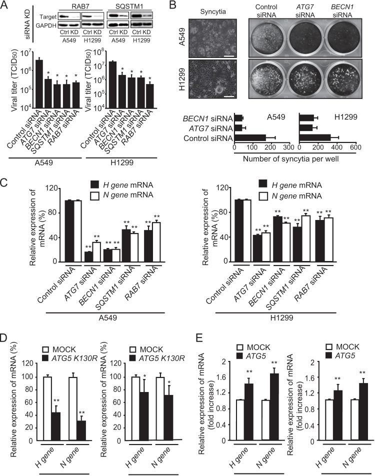
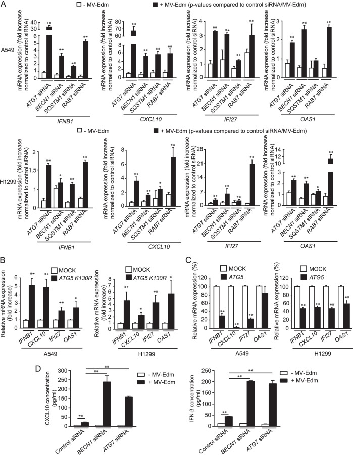
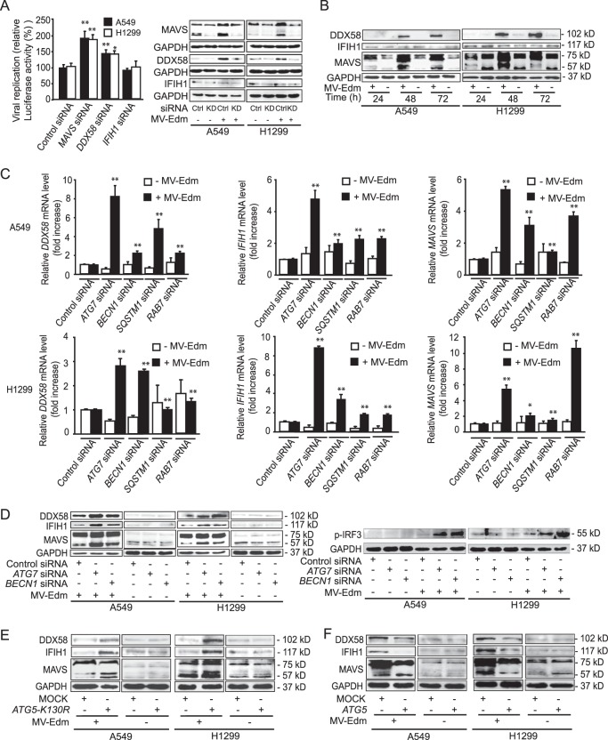
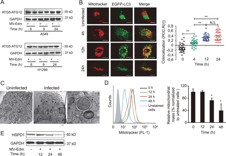
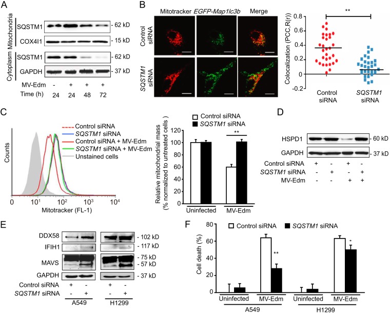
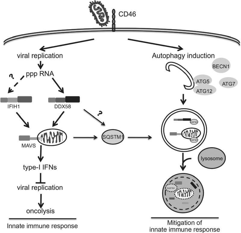
The success of future clinical trials with oncolytic viruses depends on the identification and the control of mechanisms that modulate their therapeutic efficacy. In particular, little is known about the role of autophagy in infection by attenuated measles virus of the Edmonston strain (MV-Edm). We investigated the interaction between autophagy, innate immune response, and oncolytic activity of MV-Edm, since the antiviral immune response is a known factor limiting virotherapies. We report that MV-Edm exploits selective autophagy to mitigate the innate immune response mediated by DDX58/RIG-I like receptors (RLRs) in non-small cell lung cancer (NSCLC) cells. Both RNA interference (RNAi) and overexpression approaches demonstrate that autophagy enhances viral replication and inhibits the production of type I interferons regulated by RLRs. We show that MV-Edm unexpectedly triggers SQSTM1/p62-mediated mitophagy, resulting in decreased mitochondrion-tethered mitochondrial antiviral signaling protein (MAVS) and subsequently weakening the innate immune response. These results unveil a novel infectious strategy based on the usurpation of mitophagy leading to mitigation of the innate immune response. This finding provides a rationale to modulate autophagy in oncolytic virotherapy.

Importance: In vitro studies, preclinical experiments in vivo, and clinical trials with humans all indicate that oncolytic viruses hold promise for cancer therapy. Measles virus of the Edmonston strain (MV-Edm), which is an attenuated virus derived from the common wild-type measles virus, is paradigmatic for therapeutic oncolytic viruses. MV-Edm replicates preferentially in and kills cancer cells. The efficiency of MV-Edm is limited by the immune response of the host against viruses. In our study, we revealed that MV-Edm usurps a homeostatic mechanism of intracellular degradation of mitochondria, coined mitophagy, to attenuate the innate immune response in cancer cells. This strategy might provide a replicative advantage for the virus against the development of antiviral immune responses by the host. These findings are important since they may not only indicate that inducers of autophagy could enhance the efficacy of oncolytic therapies but also provide clues for antiviral therapy by targeting SQSTM1/p62-mediated mitophagy.


Figures

FIG 1
FIG 1
MV-Edm-induced autophagy and autophagic flux enhance oncolytic activity of the virus in NSCLC cells. (A) Translocation of EGFP-MAP1LC3B toward autophagosomes was followed by fluorescence microscopy in A549 cells transiently transfected with a plasmid encoding EGFP-MAP1LC3B grown for 2 h in the presence or absence of MV-Edm (MOI = 0.5). Bars represent 10 μm. The number of EGFP-MAP1LC3B-positive vesicles per cell was quantified by fluorescence microscopy. Bright punctuate structures are GFP+ vesicles, indicating autophagosomes. (B) A549 lung cancer cells were transiently transfected with a plasmid encoding EGFP-MAP1LC3B for 24 h followed by infection with MV-Edm (MOI = 0.5) for 6 and 24 h or were left uninfected and cultured in completed medium or in Dulbecco's PBS (DPBS) for 4 h. Cells were then stained for measles virus H protein. Aggregation of EGFP-MAP1LC3B at autophagosomes (green dots) and expression of measles virus H protein (red dots) were evaluated by fluorescence confocal microscopy. Scale bars represent 10 μm. (C) Levels of lipidated MAP1LC3B (LC3-II) were assessed by Western blotting in lysates obtained from A549 and H1299 lung cancer cells infected with MV-Edm at an MOI of 0.5 or left uninfected for 6, 9, and 24 h (upper panels). The LC3-II/GAPDH ratio was quantified by densitometric analysis (lower panels). (D) MAP1LC3B lipidation was analyzed and quantified in A549 and H1299 cells infected with MV-Edm and grown with or without chloroquine. (E) Degradation of SQSTM1 was monitored by immunoblotting of A549 and H1299 cells after infection by MV-Edm (MOI = 0.2) at 24, 48, and 72 h. (F and G) A549 and H1299 cells transfected with siRNA targeting ATG5, ATG7, or BECN1 or with nontargeting control siRNA (F) or with ATG5 or ATG7 expression plasmids or a mock plasmid (G) for 24 h were infected with MV-Edm (MOI = 0.2) for 48 h. Cell death was quantified using trypan blue staining. Knockdown efficiency for ATG5, ATG7, and BECN1 was monitored at the protein level by Western blotting (F, upper panels). One experiment representative of three (for F) or of two (for G) is shown. Results are means of triplicates. (H) Uninfected A549 and H1299 cells were transfected with plasmids encoding ATG5 or ATG7 or with a mock plasmid for 19 h. Cells were then grown in the presence or absence of chloroquine (CQ) (20 μM) for another 5 h. LC3-II was evaluated by Western blotting. The LC3-II/GAPDH ratio was quantified by densitometry. *, P < 0.05; **, P < 0.01.
FIG 2
FIG 2
MV-Edm-induced autophagy and autophagic flux promote viral replication and spread in NSCLC cells. (A) Replication of MV-Edm was quantified in A549 and H1299 cells transfected with siRNA targeting ATG7, BECN1, SQSTM1, or RAB7 or with control siRNA, followed by MV-Edm infection at an MOI of 0.2 for 48 h. Viral particles were then harvested by two rounds of freezing-thawing cycles, and the viral titer was determined by calculating the TCID50 on Vero cells. Knockdown efficiency of RAB7 and SQSTM1 were monitored by Western blotting (upper panels). (B) Syncytium formation was observed by phase-contrast microscopy (left panel) and was further evaluated by crystal violet staining of A549 and H1299 cells transfected with siRNAs targeting ATG7 or BECN1 or with control siRNA and infected with MV-Edm (MOI = 0.2) for 48 h (right panel). Pictures representative of three independent experiments are shown. The mean number of syncytia is depicted (lower panel). (C to E) The expression of H and N viral structural genes was quantified by qRT-PCR in A549 and H1299 cells transfected with siRNA targeting ATG7, BECN1, SQSTM1, RAB7, or control siRNA (C) or with a plasmid expressing a mutant ATG5 gene encoding the K130R substitution (ATG5 K130R) (D) or functional ATG5 (E), followed by infection with MV-Edm (MOI = 0.2) for another 48 h. Results are mean of quadruplicates. *, P < 0.05; **, P < 0.01. Similar results were obtained in two independent experiments.
FIG 3
FIG 3
MV-Edm-induced autophagy and autophagic flux impair antiviral immune responses. Gene expression of the antiviral cytokines IFNB1, CXCL10, OAS1, and IFI27 was quantified by qRT-PCR in A549 and H1299 cells transfected with siRNAs targeting ATG7, BECN1, SQSTM1, or RAB7 or with control siRNA (A), with an expression plasmid encoding the ATG5 mutant ATG5-K130R (B), or with functional ATG5 (C) and grown in the absence or presence of MV-Edm (MOI = 0.2) for 48 h. The fold increase in gene expression shown in panel A was normalized to levels for cells treated with control siRNA in the absence (open bars) or presence (filled bars) of MV-Edm. P values were obtained by comparison with results for cells transfected with control siRNA after MV-Edm infection. The percentage decrease in gene expression shown in panel B was compared to results for cells expressing ATG5-K130R and infected with MV-Edm. Means + SD for quadruplicates are shown. Similar results were obtained in two independent experiments. (D) CXCL10 and IFNB1 were quantified by ELISA in the supernatants of A549 cells transfected with siRNAs for ATG7, BECN1, or control siRNA followed by infection with MV-Edm for 48 h (filled bars) or left uninfected (open bars). Means + SD of two experiments are shown. *, P < 0.05; **, P < 0.01.
FIG 4
FIG 4
MV-Edm-induced autophagy and autophagic flux mitigate DDX58/IFIH1/MAVS signaling. (A) Viral replication was quantified in A549 and H1299 cells transfected with siRNAs targeting DDX58, IFIH1, or MAVS or with nonspecific control siRNA after infection with MV-Edm-Luc (MOI = 0.2) for 24 h. Luciferase activity as a viral replication indicator was determined relative to that of MV-Edm-infected cells transfected with control siRNA. Results are compared to those for uninfected cells. Means + SD of quadruplicates are shown (left panel). Knockdown efficiency of MAVS, DDX58, and IFIH1 in NSCLC cells with or without MV-Edm infection was monitored by Western blot 48 h after siRNA treatment (right panel). Similar results were obtained in two independent experiments. *, P < 0.05; **, P < 0.01. (B) Expression of DDX58, IFIH1, and MAVS was determined by immunoblotting in A549 and H1299 cells infected with MV-Edm (MOI = 0.2) for 48 h. (C) Gene expression of DDX58, IFIH1, and MAVS was quantified by qRT-PCR in A549 and H1299 cells transfected with siRNAs targeting ATG7, BECN1, SQSTM1, or RAB7 or with nonspecific control siRNA and grown in the absence or presence of MV-Edm (MOI = 0.2) for 48 h. The fold increase in gene expression was normalized to results for cells treated with nonspecific control siRNA in the absence (open bars) or presence (filled bars) of MV-Edm. Means + SD of quadruplicates are shown. P values were obtained by comparison with results for cells transfected with control siRNA upon MV-Edm infection. *, P < 0.05; **, P < 0.01. Similar results were obtained in three independent experiments. (D) DDX58, IFIH1, and MAVS (left panel) and p-IRF3 (right panel) protein levels were evaluated by immunoblotting of cell lysates harvested from A549 and H1299 cells transfected with siRNAs targeting ATG7 or BECN1 or with a nonspecific control siRNA followed by MV-Edm infection (MOI = 0.2) for 48 h. A representative result from two independent experiments is shown. (E and F) A549 and H1299 cells were transiently transfected with a plasmid encoding ATG5-K130R (E) or ATG5 (F) and cultured in the presence or absence of MV-Edm (MOI = 0.2) for 48 h. Cell lysates were harvested, and DDX58, IFIH1, and MAVS protein levels were evaluated by immunoblotting. A representative result from two independent experiments is shown.
FIG 5
FIG 5
MV-Edm induces mitophagy leading to mitochondrial degradation. (A) Levels of ATG5-ATG12 conjugates were assessed by Western blotting in lysates obtained from A549 and H1299 lung cancer cells infected with MV-Edm at an MOI of 0.5 or left uninfected for 6, 9, and 24 h. A representative result from two independent experiments is shown. (B) Colocalization of autophagosomes and mitochondria was quantified in A549 cells transiently transfected with pBABEpuro-EGFP-Map1lc3b and infected by MV-Edm (MOI = 0.5) for 4, 12, and 24 h or left uninfected. Cells were then stained with MitoTracker stain and subjected to confocal microscopy (left panels). Scale bars = 10 μm. Colocalization (yellow dots) of mitochondria (red) with autophagosomes (green puncta) was quantified by calculating Pearson's correlation coefficient [PCC, R(r)] (right panels). Means are shown (n = 30 for each time point). **, P < 0.01; N.S, not significant. (C) Subcellular analysis of A549 cells infected without (left panel) or with (middle and right panels) MV-Edm (MOI = 1; 24 h) was performed by electron microscopy. Arrowheads depict double-layer structures that contain mitochondria in an MV-Edm-infected cell. Scale bar = 2 μm (left), 5 μm (middle), or 1 μm (right). (D) Mitochondrial mass was measured by cytometry in MitoTracker green-stained A549 cells 12, 24, and 48 h after infection with MV-Edm (MOI = 0.5). An overlay of histograms representative of 3 independent experiments (left panel), and quantification of mitochondrial mass as mean fluorescence intensity averaged from 3 independent experiments (right panel) are shown. (E) Mitochondrial HSPD1 protein level was determined by Western blotting in lysates obtained from A549 lung cancer cells infected with MV-Edm at an MOI of 0.5 for 12, 24, and 48 h or left uninfected. A representative result from two independent experiments is shown.
FIG 6
FIG 6
SQSTM1 mediates MV-Edm-induced mitophagy, contributing to impaired DDX58/IFIH1/MAVS signaling. (A) Mitochondria were fractionated from A549 cells that were not infected or that were infected by MV-Edm at an MOI of 0.2 for 24, 48, and 72 h. Mitochondrion-associated and cytoplasmic SQSTM1 was detected by immunoblotting. COX4I1 and GAPDH were used as loading controls for mitochondrial and cytoplasmic samples, respectively. One representative blot from two independent experiments is shown. (B) Colocalization of autophagosomes and mitochondria was quantified in A549 cells transfected with SQSTM1 siRNA for 24 h followed by transient transfection with pEGFP-Map1lc3b for another 24 h. Cells were then infected with MV-Edm (MOI = 0.5) for 12 h and stained with MitoTracker red before being subjected to confocal microscopy (left panel). Scale bars = 10 μm. Colocalization (yellow dots) of mitochondria (red) with autophagosomes (green puncta) was quantified by calculating Pearson's correlation coefficient [PCC, R(r)] (right panel). Means are shown (n = 30 for each group). (C) Mitochondrial mass was measured by cytometry in MitoTracker green-stained A549 cells transfected with siRNA targeting SQSTM1 or nonspecific control siRNA and cultured in the presence or absence of MV-Edm (MOI = 0.5) for 48 h. An overlay of histograms representative of 3 independent experiments (left panel) and quantification of mitochondrial mass as mean fluorescence intensity averaged from 3 independent experiments (right panel) are shown. (D) The mitochondrial HSPD1 protein level was determined by Western blotting in lysates obtained from A549 lung cancer cells transfected with siRNA targeting SQSTM1 or nonspecific control siRNA and cultured in the presence or absence of MV-Edm (MOI = 0.5) for 48 h. A representative result from two independent experiments is shown. (E) Expression levels of DDX58, IFIH1, and MAVS were determined by immunoblotting in cell lysates from A549 and H1299 cells transfected with siRNA targeting SQSTM1 or with a nontargeting siRNA followed by infection with MV-Edm (MOI = 0.2) for 48 h. Similar results were obtained in two independent experiments. (F) Cell death was quantified by trypan blue exclusion in A549 and H1299 cells transfected with siRNA targeting SQSTM1 or with nontargeting control siRNA for 24 h and cultured in the presence or absence of MV-Edm (MOI = 0.2) for another 48 h. Means + SD for triplicates are shown; *, P < 0.05; **, P < 0.01. Similar results were obtained in three independent experiments.
FIG 7
FIG 7
MV-Edm subverts mitophagy to attenuate the innate immune response. In this model, MV-Edm induces an innate immune response via activation of DDX58/MAVS signaling while in parallel stimulating autophagy and mitophagy that degrade DDX58, IFIH1, and mitochondrion-anchored MAVS. SQSTM1 may play a crucial role in tipping the balance of anti- to proviral functions of autophagy, enhancing replication of MV-Edm, since it mediates mitophagy.

References

    1. Roy CR, Mocarski ES. 2007. Pathogen subversion of cell-intrinsic innate immunity. Nat. Immunol. 8:1179–1187. 10.1038/ni1528 - DOI - PubMed
    1. Kawai T, Takahashi K, Sato S, Coban C, Kumar H, Kato H, Ishii KJ, Takeuchi O, Akira S. 2005. IPS-1, an adaptor triggering RIG-I- and Mda5-mediated type I interferon induction. Nat. Immunol. 6:981–988. 10.1038/ni1243 - DOI - PubMed
    1. Lou Y-J, Zhang Z-L, Pan X-R, Xu G-P, Jia P-M, Li D, Tong J-H. 2011. Intact JAK-STAT signaling pathway is a prerequisite for STAT1 to reinforce the expression of RIG-G gene. Exp. Cell Res. 317:513–520. 10.1016/j.yexcr.2010.10.025 - DOI - PubMed
    1. Saito T, Hirai R, Loo Y-M, Owen D, Johnson CL, Sinha SC, Akira S, Fujita T, Gale M. 2007. Regulation of innate antiviral defenses through a shared repressor domain in RIG-I and LGP2. Proc. Natl. Acad. Sci. U. S. A. 104:582–587. 10.1073/pnas.0606699104 - DOI - PMC - PubMed
    1. Hall JC, Rosen A. 2010. Type I interferons: crucial participants in disease amplification in autoimmunity. Nat. Rev. Rheumatol. 6:40–49. 10.1038/nrrheum.2009.237 - DOI - PMC - PubMed

Publication types

LinkOut - more resources