

Pathophysiology of merkel cell
- PMID: 24574661
- PMCID: PMC3927344
- DOI: 10.4103/0973-029X.125208
Pathophysiology of merkel cell
Abstract
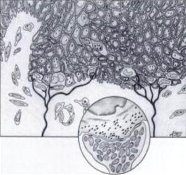
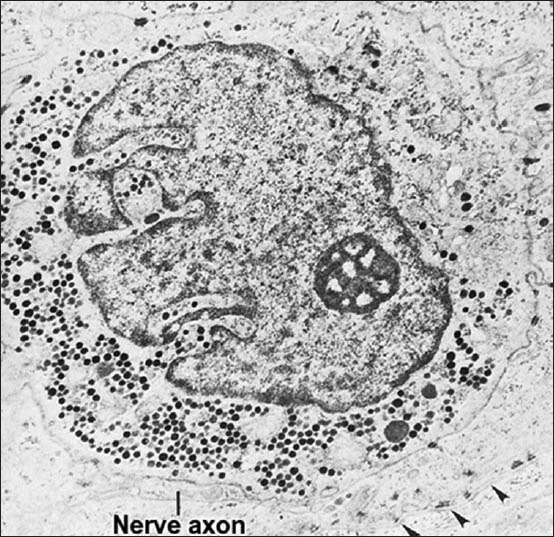
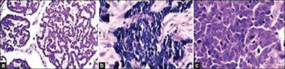
The objective of this review is to introduce Merkel cells (MCs), to provide a basic overview on the theoretical background of function, development and clinical importance of MCs. The origin of human MCs have been controversial. Some investigators believe that it is a neural crest derivate, whereas others have proposed that it is a differentiation product of the fetal epidermal keratinocytes. MCs are cells primarily localized in the epidermal basal layer of vertebrates and concentrated in touch-sensitive areas in glabrous, hairy skin and in some mucosa. In routine light microscopy, human MCs can hardly be identified. Cytokeratin 20 (CK20) is a reliable marker with highest degree of specificity. MCs can be also distinguished by electron microscopy. MC carcinoma (MCC) is an uncommon and often aggressive malignancy and found mainly in elderly patients. It occurs most frequently in the head and neck region. Diagnosis is based on typical histological presentation on hematoxylin and eosin (H and E) stained slides together with the results of immunohistochemistry. Histologically, MCC has been classified into three distinct subtypes: Trabecular, intermediate and small cell type.
Keywords: Cytokeratin 20; merkel cell carcinoma; merkel cells.
Conflict of interest statement
Figures
References
-
- Tachibana T. The Merkel cell: Recent findings and unresolved problems. Arch Histol Cytol. 1995;58:379–96. - PubMed
-
- Tweedle CD. Ultrastructure of Merkel cell development in aneurogenic and control amphibian larvae (ambystoma) Neuroscience. 1978;3:481–6. - PubMed
-
- Moll I, Lane AT, Franke WW, Moll R. Intra epidermal formation of Merkel cell in xenografts of human foetal skin. J Invest Dermatol. 1990;94:359–64. - PubMed
-
- Grim M, Halata Z. Developmental origin of avian Merkel cell. Anat Embryol (Berl) 2000;202:401–10. - PubMed
-
- Halata Z, Grim M, Christ B. Origin of spinal cord meninges sheaths of peripheral nerves, and cutaneous receptors including Merkel cell. An experimental and ultrastructural study with avian chimeras. Anat Embryol (Berl) 1990;182:529–37. - PubMed
Publication types
LinkOut - more resources
Full Text Sources
Other Literature Sources
