

Myoepithelial carcinoma arising in recurrent pleomorphic adenoma in maxillary sinus
- PMID: 24574666
- PMCID: PMC3927349
- DOI: 10.4103/0973-029X.125213
Myoepithelial carcinoma arising in recurrent pleomorphic adenoma in maxillary sinus
Abstract
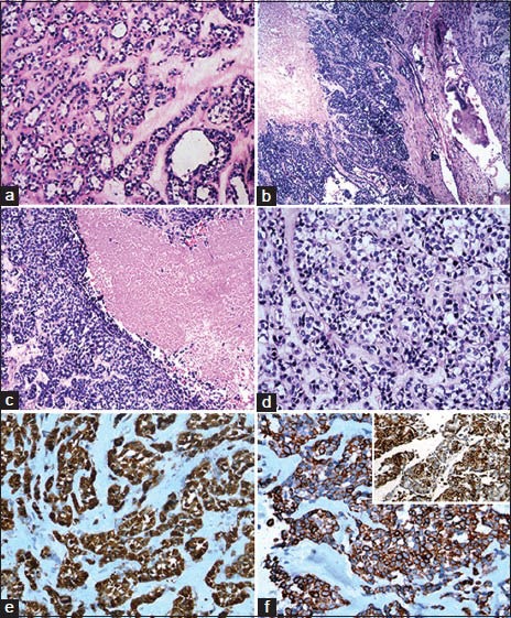
Myoepithelial carcinoma is characterized by nearly exclusive myoepithelial differentiation and evidence of malignancy. It may arise de novo or in preexisting benign tumors including pleomorphic adenoma and benign myoepithelioma. A 39-year-old lady presented with painless progressive swelling on the right cheek and right side of palate. On surgery, there was a mass in right maxillary sinus which was surgically excised and diagnosed on histopathology as pleomorphic adenoma. Subsequently, there were two recurrences. The first recurrence was in the right maxilla after 2 years that was removed surgically and diagnosed as pleomorphic adenoma. One year later, she came with rapidly progressive swelling in bilateral cheeks and face. Intraoperatively, there was a large tumor in both maxillary sinuses with extensive local infiltration. Histologically, it was diagnosed as myoepithelial carcinoma. Carcinoma ex pleomorphic adenoma is usually a high grade malignancy. It occurs most commonly in parotid gland followed by submandibular glands, minor salivary glands and occasionally in sublingual gland. To the best of our knowledge, this is the first case of myoepithelial carcinoma arising in a recurrent pleomorphic adenoma in the maxillary sinus.
Keywords: Carcinoma ex-pleomorphic adenoma; maxillary sinus; myoepithelial carcinoma.
Conflict of interest statement
Figures
References
-
- Wenig BM, Heffer CS, editors. 2nd ed. Philadelphia: WB Saunders; 2008. Atlas of Head and Neck Pathology; pp. 582–702.
-
- Lewis EJ, Olsen KD, Sebo TJ. Carcinoma ex pleomorphic adenoma: Pathological analysis of 73 cases. Hum Pathol. 2001;32:596–604. - PubMed
-
- Shin SY, Park JG, Ahn ST. A case of carcinoma ex pleomorphic adenoma in the maxillary sinus. J Korean Soc Plast Reconstr Surg. 2001;28:421–3.
-
- Barnes L, Eveson JW, Reichart P, Sidransky D, editors. Lyon, France: IARC Press; 2005. Pathology and Genetics of Head and Neck Tumours. World Health Organisation Classification of Tumours.
Publication types
LinkOut - more resources
Full Text Sources
Other Literature Sources
