

A completely isolated intestinal duplication cyst mimicking ovarian cyst torsion in an adult
- PMID: 24574732
- PMCID: PMC3923038
- DOI: 10.3748/wjg.v20.i2.603
A completely isolated intestinal duplication cyst mimicking ovarian cyst torsion in an adult
Abstract
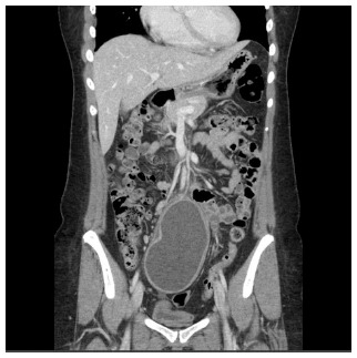
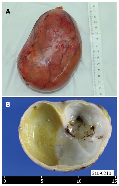
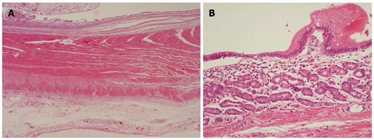
Intestinal duplications are rare congenital anomalies that can occur anywhere in the gastrointestinal tract. They are most commonly located in the ileum and are usually detected in infancy or early childhood. Duplicated segments are usually firmly attached to and sometimes communicate with the normal gastrointestinal tract. Rarely, intestinal duplications are completely isolated, thus not associated at all with any part of the gastrointestinal tract. Such duplications do not share a common blood supply with the adjacent normal intestinal segment, unlike the usual form of duplication, but rather have a separate vascular pedicle. Reports of completely isolated duplication cysts in adults are extremely rare; we found only five such reports in the English-language medical literature. Here, we report a case of a completely isolated duplication cyst 12 cm long in an adult female. The cyst had no connection to any part of the intestinal tract and had a dedicated vascular pedicle.
Keywords: Adult; Congenital abnormalities; Cysts; Digestive system; Duplication.
Figures
Similar articles
-
Noncommunicating isolated enteric duplication cyst in childhood.J Pediatr Surg. 2009 Jul;44(7):e9-e10. doi: 10.1016/j.jpedsurg.2009.03.041. J Pediatr Surg. 2009. PMID: 19573650
-
Duplication cyst of ileum presenting as acute intestinal obstruction in an adult.BMJ Case Rep. 2016 Oct 6;2016:bcr2016214775. doi: 10.1136/bcr-2016-214775. BMJ Case Rep. 2016. PMID: 27758850 Free PMC article.
-
Isolated enteric duplication cyst with respiratory epithelium: case report and review of the literature.Eur J Pediatr Surg. 2008 Oct;18(5):337-9. doi: 10.1055/s-2008-1038646. Epub 2008 Oct 15. Eur J Pediatr Surg. 2008. PMID: 18924072
-
An unusual presentation of intestinal duplication with a literature review.Dig Dis Sci. 1996 Mar;41(3):627-9. doi: 10.1007/BF02282353. Dig Dis Sci. 1996. PMID: 8617148 Review.
-
Perforated ileal duplication cyst with haemorrhagic pseudocyst formation.Pediatr Radiol. 2003 Jul;33(7):489-91. doi: 10.1007/s00247-003-0880-2. Epub 2003 Apr 24. Pediatr Radiol. 2003. PMID: 12712267 Review.
Cited by
-
Laparoscopic excision of a retroperitoneal completely isolated enteric duplication cyst in an adult male: A case report and review of literature.Int J Surg Case Rep. 2018;46:1-5. doi: 10.1016/j.ijscr.2018.03.035. Epub 2018 Mar 28. Int J Surg Case Rep. 2018. PMID: 29626802 Free PMC article.
-
Two Cases of Adult-Onset Intestinal Duplication Manifested as Acute Abdomen: Case Report and Review of the Literature.Surg Case Rep. 2025;11(1):24-0023. doi: 10.70352/scrj.cr.24-0023. Epub 2025 Feb 6. Surg Case Rep. 2025. PMID: 40008372 Free PMC article.
-
Laparoscopic approach to non-communicating intestinal duplication cyst in adult.J Surg Case Rep. 2018 Apr 3;2018(4):rjy061. doi: 10.1093/jscr/rjy061. eCollection 2018 Apr. J Surg Case Rep. 2018. PMID: 29644042 Free PMC article.
-
A huge completely isolated duplication cyst complicated by torsion and lined by 3 different mucosal epithelial components in an adult: A case report.Medicine (Baltimore). 2018 Nov;97(44):e13005. doi: 10.1097/MD.0000000000013005. Medicine (Baltimore). 2018. PMID: 30383655 Free PMC article.
-
Report of a rare case and review of adult intestinal duplication at the opposite side of mesenteric margin.Sao Paulo Med J. 2018 Jan-Feb;136(1):89-93. doi: 10.1590/1516-3180.2017.0184030817. Epub 2017 Dec 7. Sao Paulo Med J. 2018. PMID: 29236936 Free PMC article. Review.
References
-
- Macpherson RI. Gastrointestinal tract duplications: clinical, pathologic, etiologic, and radiologic considerations. Radiographics. 1993;13:1063–1080. - PubMed
-
- Sinha A, Ojha S, Sarin YK. Completely isolated, noncontiguous duplication cyst. Eur J Pediatr Surg. 2006;16:127–129. - PubMed
-
- Gilbert-Barness E. Potter’s pathology of the fetus, infant and child. 2nd ed. Amsterdam, Paesi Bassi: Elsevier Inc; 2007. pp. 1169–1170.
Publication types
MeSH terms
LinkOut - more resources
Full Text Sources
Other Literature Sources
Medical
