

A case report of anaplastic carcinoma of the pancreas with remarkable intraductal tumor growth into the main pancreatic duct
- PMID: 24574758
- PMCID: PMC3921494
- DOI: 10.3748/wjg.v20.i3.852
A case report of anaplastic carcinoma of the pancreas with remarkable intraductal tumor growth into the main pancreatic duct
Abstract
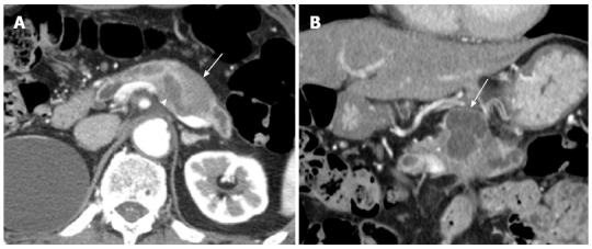
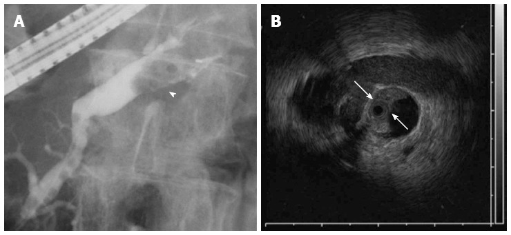
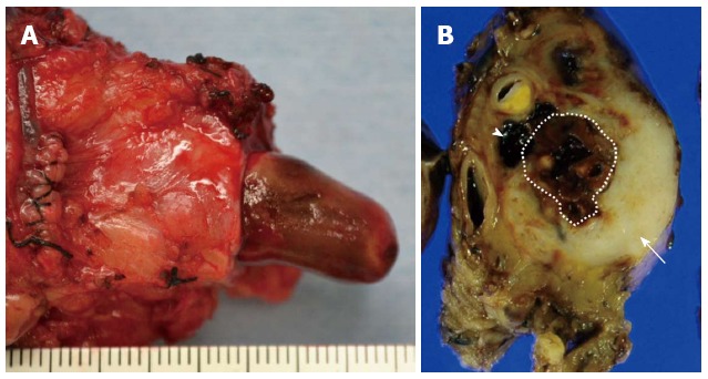
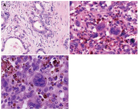
We herein report a case of anaplastic carcinoma of the pancreas with remarkable intraductal tumor growth into the main pancreatic duct. A 76-year-old male was referred to our hospital for treatment of a pancreatic tumor. Preoperative examinations revealed a poorly defined tumor in the main pancreatic duct in the body of the pancreas, accompanied with severe dilatation of the main pancreatic duct, which was diagnosed as an intraductal papillary-mucinous neoplasm. We performed distal pancreatectomy and splenectomy. The pathological examination revealed that the tumor consisted of a mixture of anaplastic carcinoma (giant cell type) and adenocarcinoma in the pancreas. There was a papillary projecting tumor composed of anaplastic carcinoma in the dilated main pancreatic duct. The patient is now receiving chemotherapy because liver metastasis was detected 12 mo after surgery. In this case, we could observe a remarkable intraductal tumor growth into the main pancreatic duct. We also discuss the pathogenesis and characteristics of this rare tumor with specific tumor growth.
Keywords: Anaplastic carcinoma; Giant cell carcinoma; Intraductal tumor growth; Papillary projecting tumor.
Figures

Similar articles
-
Synchronous pancreatic solid pseudopapillary neoplasm and intraductal papillary mucinous neoplasm.World J Gastroenterol. 2013 Jun 7;19(21):3358-63. doi: 10.3748/wjg.v19.i21.3358. World J Gastroenterol. 2013. PMID: 23745041 Free PMC article. Review.
-
Partial pancreatic head resection for intraductal papillary mucinous carcinoma originating in a branch of the duct of santorini.Eur Surg Res. 2002 Nov-Dec;34(6):437-40. doi: 10.1159/000065707. Eur Surg Res. 2002. PMID: 12403944
-
[A case report of total remnant pancreatectomy for ductal carcinoma after distal pancreatectomy for invasive intraductal papillary mucinous carcinoma].Gan To Kagaku Ryoho. 2012 Nov;39(12):2140-2. Gan To Kagaku Ryoho. 2012. PMID: 23268003 Japanese.
-
Sequential progression and intraductal spread of invasive ductal adenocarcinoma of the pancreas arising from around the main pancreatic duct.Hepatogastroenterology. 2005 May-Jun;52(63):745-8. Hepatogastroenterology. 2005. PMID: 15966196
-
Intraductal papillary mucinous carcinoma of the pancreas associated with pancreas divisum: a case report and review of the literature.BMC Gastroenterol. 2015 Jul 8;15:78. doi: 10.1186/s12876-015-0313-3. BMC Gastroenterol. 2015. PMID: 26152300 Free PMC article. Review.
Cited by
-
A case of undifferentiated carcinoma of the pancreas mimicking main-duct intraductal papillary mucinous neoplasm (IPMN).Abdom Imaging. 2015 Mar;40(3):466-70. doi: 10.1007/s00261-014-0326-3. Abdom Imaging. 2015. PMID: 25526684 Free PMC article.
-
An Autopsy Case of Anaplastic Pancreatic Ductal Carcinoma (Spindle Cell Type) Multiple Onset in the Pancreas.Case Rep Oncol. 2019 Apr 24;12(1):332-338. doi: 10.1159/000499969. eCollection 2019 Jan-Apr. Case Rep Oncol. 2019. PMID: 31123460 Free PMC article.
-
Rapidly Progressing Anaplastic Carcinoma of the Pancreas with Mucoepidermoid Carcinoma: An Autopsy Case Report.Intern Med. 2021 Jul 15;60(14):2235-2240. doi: 10.2169/internalmedicine.6181-20. Epub 2021 Feb 22. Intern Med. 2021. PMID: 33612673 Free PMC article.
-
Anaplastic Carcinoma of the Pancreas: A Rare Clinical Entity.Cureus. 2017 Oct 18;9(10):e1782. doi: 10.7759/cureus.1782. Cureus. 2017. PMID: 29279808 Free PMC article.
-
Cephalic undifferentiated carcinoma with osteoclast-like giant cells arising from the main pancreatic duct: case report and literature review.Arch Clin Cases. 2021 Oct 27;6(1):6-21. doi: 10.22551/2019.22.0601.10148. eCollection 2019. Arch Clin Cases. 2021. PMID: 34754903 Free PMC article.
References
-
- Chen J, Baithun SI. Morphological study of 391 cases of exocrine pancreatic tumours with special reference to the classification of exocrine pancreatic carcinoma. J Pathol. 1985;146:17–29. - PubMed
-
- Ichikawa T, Federle MP, Ohba S, Ohtomo K, Sugiyama A, Fujimoto H, Haradome H, Araki T. Atypical exocrine and endocrine pancreatic tumors (anaplastic, small cell, and giant cell types): CT and pathologic features in 14 patients. Abdom Imaging. 2000;25:409–419. - PubMed
-
- Paal E, Thompson LD, Frommelt RA, Przygodzki RM, Heffess CS. A clinicopathologic and immunohistochemical study of 35 anaplastic carcinomas of the pancreas with a review of the literature. Ann Diagn Pathol. 2001;5:129–140. - PubMed
-
- Strobel O, Hartwig W, Bergmann F, Hinz U, Hackert T, Grenacher L, Schneider L, Fritz S, Gaida MM, Büchler MW, et al. Anaplastic pancreatic cancer: Presentation, surgical management, and outcome. Surgery. 2011;149:200–208. - PubMed
-
- Clark CJ, Graham RP, Arun JS, Harmsen WS, Reid-Lombardo KM. Clinical outcomes for anaplastic pancreatic cancer: a population-based study. J Am Coll Surg. 2012;215:627–634. - PubMed
Publication types
MeSH terms
LinkOut - more resources
Full Text Sources
Other Literature Sources
Medical
