Novel esophageal squamous cell carcinoma bone metastatic clone isolated by scintigraphy, X ray and micro PET/CT
- PMID: 24574775
- PMCID: PMC3921526
- DOI: 10.3748/wjg.v20.i4.1030
Novel esophageal squamous cell carcinoma bone metastatic clone isolated by scintigraphy, X ray and micro PET/CT
Abstract
Aim: To establish a Chinese esophageal squamous cell carcinoma (ESCC) cell line with high bone metastasis potency using (99m)Tc-methylene diphosphonate ((99m)Tc-MDP) micro-pinhole scintigraphy, X ray and micro-positron emission tomography/computed tomography (PET/CT) for exploring the mechanism of occurrence and development in esophageal cancer.
Methods: The cells came from a BALB/c nu/nu immunodeficient mouse, and oncogenic tumor tissue was from a surgical specimen from a 61-year-old male patient with ESCC. The cell growth curve was mapped and analysis of chromosome karyotype was performed. Approximately 1 × 10⁶ oncogenic cells were injected into the left cardiac ventricle of immunodeficient mice. The bone metastatic lesions of tumor-bearing mice were detected by (99m)Tc-MDP scintigraphy, micro-PET/CT and X-ray, and were resected from the mice under deep anesthesia. The bone metastatic cells in the lesions were used for culture and for repeated intracardiac inoculation. This in vivo/in vitro experimental metastasis study was repeated for four cycles. All of the suspicious bone sites were confirmed by pathology. Real-time polymerase chain reaction was used to compare the gene expression in the parental cells and in the bone metastatic clone.
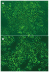
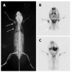
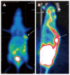
Results: The surgical specimen was implanted subcutaneously in immunodeficient mice and the tumorigenesis rate was 100%. First-passage oncogenic cells were named CEK-Sq-1. The chromosome karyotype analysis of the cell line was hypotriploid. The bone metastasis rate went from 20% with the first-passage oncogenic cells via intracardiac inoculation to 90% after four cycles. The established bone metastasis clone named CEK-Sq-1BM had a high potential to metastasize in bone, including mandible, humerus, thoracic and lumbar vertebrae, scapula and femur. The bone metastasis lesions were successfully detected by micro-pinhole bone scintigraphy, micro-PET/CT, and X-ray. The sensitivity, specificity and accuracy of the micro-pinhole scintigraphy, X-ray, and micro-PET/CT imaging examinations were: 89.66%/32%/80%, 88.2%/100%/89.2%, and 88.75%/77.5%/87.5%, respectively. Some gene expression difference was found between parental and bone metastasis cells.
Conclusion: This newly established Chinese ESCC cell line and animal model may provide a useful tool for the study of the pathogenesis and development of esophageal carcinoma.
Keywords: Bone metastasis; Cell line; Esophageal squamous cell carcinoma; Molecular imaging; Real-time polymerase chain reaction.
Figures


Similar articles
-
Establishment of an experimental human lung adenocarcinoma cell line SPC-A-1BM with high bone metastases potency by (99m)Tc-MDP bone scintigraphy.Nucl Med Biol. 2009 Apr;36(3):313-21. doi: 10.1016/j.nucmedbio.2008.12.007. Nucl Med Biol. 2009. PMID: 19324277
-
[Establishment of a novel chinese human lung adenocarcinoma cell line CPA-Yang3 and its real bone metastasis clone CPA-Yang3BM in immunodeficient mice].Zhongguo Fei Ai Za Zhi. 2011 Feb;14(2):79-85. doi: 10.3779/j.issn.1009-3419.2011.02.01. Zhongguo Fei Ai Za Zhi. 2011. PMID: 21342638 Free PMC article. Chinese.
-
Prospective study evaluating the relative sensitivity of 18F-NaF PET/CT for detecting skeletal metastases from renal cell carcinoma in comparison to multidetector CT and 99mTc-MDP bone scintigraphy, using an adaptive trial design.Ann Oncol. 2015 Oct;26(10):2113-8. doi: 10.1093/annonc/mdv289. Epub 2015 Jul 22. Ann Oncol. 2015. PMID: 26202597 Free PMC article.
-
[Combining CT and scintigraphy: SPECT-CT and PET-CT].Ned Tijdschr Geneeskd. 2011;155(36):A2792. Ned Tijdschr Geneeskd. 2011. PMID: 21914229 Review. Dutch.
-
Evaluation of preoperative staging for esophageal squamous cell carcinoma.World J Gastroenterol. 2016 Aug 7;22(29):6683-9. doi: 10.3748/wjg.v22.i29.6683. World J Gastroenterol. 2016. PMID: 27547011 Free PMC article. Review.
Cited by
-
Frequent, quantitative bone planar scintigraphy for determination of bone anabolism in growing mice.PeerJ. 2021 Dec 9;9:e12355. doi: 10.7717/peerj.12355. eCollection 2021. PeerJ. 2021. PMID: 34966570 Free PMC article.
-
Oesophageal carcinoma with solitary patellar metastasis: a rare case report.J Int Med Res. 2021 Apr;49(4):3000605211009812. doi: 10.1177/03000605211009812. J Int Med Res. 2021. PMID: 33906528 Free PMC article.
References
-
- Jemal A, Center MM, DeSantis C, Ward EM. Global patterns of cancer incidence and mortality rates and trends. Cancer Epidemiol Biomarkers Prev. 2010;19:1893–1907. - PubMed
-
- Jemal A, Siegel R, Ward E, Hao Y, Xu J, Thun MJ. Cancer statistics, 2009. CA Cancer J Clin. 2009;59:225–249. - PubMed
-
- Siegel R, Naishadham D, Jemal A. Cancer statistics, 2012. CA Cancer J Clin. 2012;62:10–29. - PubMed
-
- Yang L, Parkin DM, Li L, Chen Y. Time trends in cancer mortality in China: 1987-1999. Int J Cancer. 2003;106:771–783. - PubMed
-
- Shimada Y, Imamura M, Wagata T, Yamaguchi N, Tobe T. Characterization of 21 newly established esophageal cancer cell lines. Cancer. 1992;69:277–284. - PubMed
Publication types
MeSH terms
Substances
LinkOut - more resources
Full Text Sources
Other Literature Sources
Medical
Research Materials
Miscellaneous

