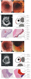
Endoscopic ultrasonography for staging of T1a and T1b esophageal squamous cell carcinoma
- PMID: 24574809
- PMCID: PMC3921517
- DOI: 10.3748/wjg.v20.i5.1340
Endoscopic ultrasonography for staging of T1a and T1b esophageal squamous cell carcinoma
Abstract
Aim: To investigate the accuracy of Endoscopic ultrasound (EUS) in staging and sub-staging T1a and T1b esophageal squamous cell carcinoma (ESCC).
Methods: A retrospective analysis involving 72 patients with pathologically confirmed T1a or T1b ESCC, was undertaken between January 2005 and December 2011 in Sun Yat-sen University Cancer Center. The accuracy and efficiency of EUS for detecting stages T1a and T1b ESCC were examined.
Results: The overall accuracy of EUS for detecting stage T1a or T1b ESCC was 70.8% (51/72), and the sensitivity was 74.3%. 77.8% (7/9) of lesions originated in the upper thoracic region, 73.1% (38/52) in the mid-thoracic region and 72.7% (8/11) in the lower thoracic region. Multivariate analysis revealed that the diagnostic accuracy of EUS was closely related to lesion length (F = 4.984, P = 0.029).
Conclusion: EUS demonstrated median degree of accuracy for distinguishing between stages T1a and T1b ESCC. Therefore, it is necessary to improve EUS for staging early ESCC.
Keywords: Early cancer; Endoscopic ultrasound; Esophageal cancer; Squamous carcinoma; Stage.
Figures

Similar articles
-
Endoscopic Ultrasound for Preoperative Esophageal Squamous Cell Carcinoma: a Meta-Analysis.PLoS One. 2016 Jul 7;11(7):e0158373. doi: 10.1371/journal.pone.0158373. eCollection 2016. PLoS One. 2016. PMID: 27387830 Free PMC article.
-
Submucosal Saline Injection Followed by Endoscopic Ultrasound versus Endoscopic Ultrasound Only for Distinguishing between T1a and T1b Esophageal Cancer.Clin Cancer Res. 2020 Jan 15;26(2):384-390. doi: 10.1158/1078-0432.CCR-19-1722. Epub 2019 Oct 15. Clin Cancer Res. 2020. PMID: 31615934 Clinical Trial.
-
Endoscopic ultrasound combined with submucosal saline injection for differentiation of T1a and T1b esophageal squamous cell carcinoma: a novel technique.Endoscopy. 2013 Aug;45(8):667-70. doi: 10.1055/s-0033-1344024. Epub 2013 Jun 27. Endoscopy. 2013. PMID: 23807801
-
Evaluation of preoperative staging for esophageal squamous cell carcinoma.World J Gastroenterol. 2016 Aug 7;22(29):6683-9. doi: 10.3748/wjg.v22.i29.6683. World J Gastroenterol. 2016. PMID: 27547011 Free PMC article. Review.
-
Role of endoscopic ultrasonography in the diagnosis of early esophageal carcinoma.Gastrointest Endosc Clin N Am. 2005 Jan;15(1):93-9, ix. doi: 10.1016/j.giec.2004.07.015. Gastrointest Endosc Clin N Am. 2005. PMID: 15555954 Review.
Cited by
-
Role of clip markers placed by endoscopic ultrasonography in contouring gross tumor volume for thoracic esophageal squamous cell carcinoma: one prospective study.Ann Transl Med. 2020 Sep;8(18):1144. doi: 10.21037/atm-20-4030. Ann Transl Med. 2020. PMID: 33240993 Free PMC article.
-
Endoscopic Ultrasound for Preoperative Esophageal Squamous Cell Carcinoma: a Meta-Analysis.PLoS One. 2016 Jul 7;11(7):e0158373. doi: 10.1371/journal.pone.0158373. eCollection 2016. PLoS One. 2016. PMID: 27387830 Free PMC article.
-
Role of endoscopic therapy in early esophageal cancer.World J Gastroenterol. 2018 Sep 21;24(35):3965-3973. doi: 10.3748/wjg.v24.i35.3965. World J Gastroenterol. 2018. PMID: 30254401 Free PMC article.
-
Update on Endoscopy-Based Imaging Techniques in the Diagnosis of Esophageal Cancer.Curr Health Sci J. 2017 Oct-Dec;43(4):295-300. doi: 10.12865/CHSJ.43.04.01. Epub 2017 Dec 28. Curr Health Sci J. 2017. PMID: 30595892 Free PMC article.
-
A deep learning-based system to identify originating mural layer of upper gastrointestinal submucosal tumors under EUS.Endosc Ultrasound. 2023 Nov-Dec;12(6):465-471. doi: 10.1097/eus.0000000000000029. Epub 2023 Dec 22. Endosc Ultrasound. 2023. PMID: 38948124 Free PMC article.
References
-
- Union for International Cancer Control (UICC) TNM Classification of Malignant Tumors. 7th ed. Oxford: Wiley-Blackwell; 2009. pp. 15–18.
-
- National Comprehensive Cancer Network (NCCN) NCCN practice guidline for oncology (esophageal and esophagogastric junction cancer) version 2. 2011. Available from: http://www.nccn.org/professionals/physician_gls/f_guidelines.asp#site.
-
- Higuchi K, Tanabe S, Koizumi W, Sasaki T, Nakatani K, Saigenji K, Kobayashi N, Mitomi H. Expansion of the indications for endoscopic mucosal resection in patients with superficial esophageal carcinoma. Endoscopy. 2007;39:36–40. - PubMed
-
- Nealis TB, Washington K, Keswani RN. Endoscopic therapy of esophageal premalignancy and early malignancy. J Natl Compr Canc Netw. 2011;9:890–899. - PubMed
Publication types
MeSH terms
LinkOut - more resources
Full Text Sources
Other Literature Sources
Medical

