Review
. 2014 Feb 11:5:20.
doi: 10.3389/fphys.2014.00020. eCollection 2014.

Connexin hemichannels in the lens

Affiliations
Review

Connexin hemichannels in the lens

Eric C Beyer et al. Front Physiol. .

Abstract

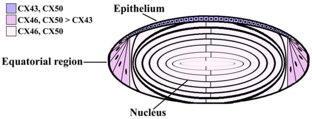
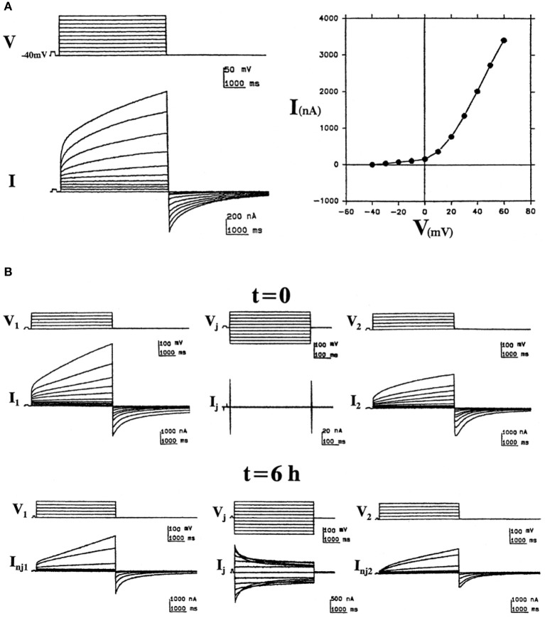
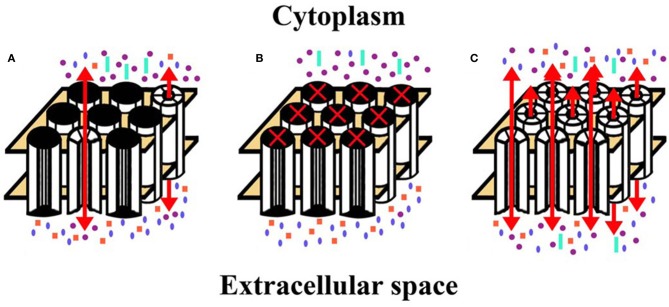
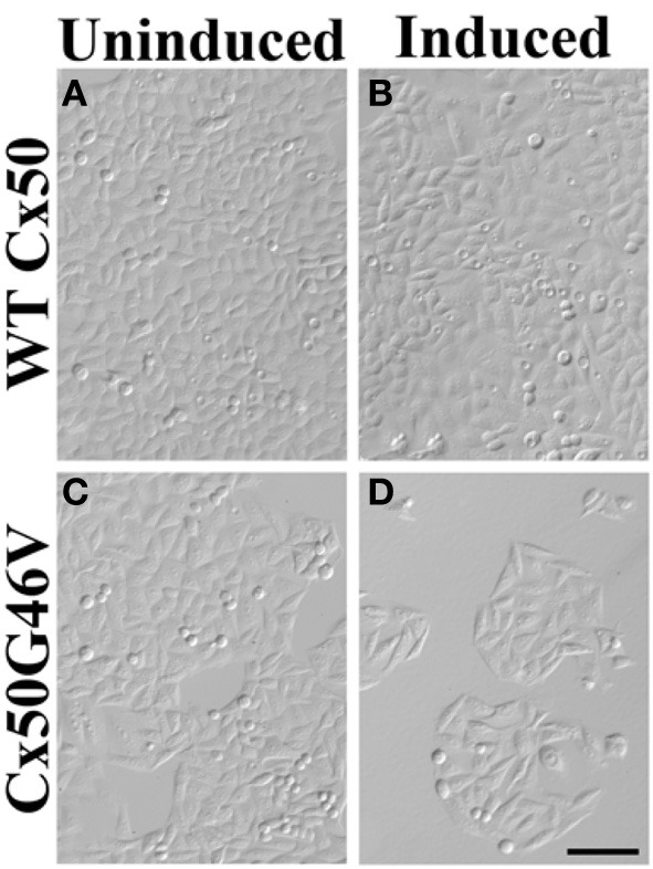
The normal function and survival of cells in the avascular lens is facilitated by intercellular communication through an extensive network of gap junctions formed predominantly by three connexins (Cx43, Cx46, and Cx50). In expression systems, these connexins can all induce hemichannel currents, but other lens proteins (e.g., pannexin1) can also induce similar currents. Hemichannel currents have been detected in isolated lens fiber cells. These hemichannels may make significant contributions to normal lens physiology and pathophysiology. Studies of some connexin mutants linked to congenital cataracts have implicated hemichannels with aberrant voltage-dependent gating or modulation by divalent cations in disease pathogenesis. Hemichannels may also contribute to age- and disease-related cataracts.

Keywords: cataract; connexin46; connexin50; gap junction; lens.


Figures

Figure 1
Figure 1
Diagram of the lens showing the distribution of connexin isoforms. Cells from the anterior epithelial cell layer express Cx43 and Cx50, differentiating fiber cells express Cx43, Cx46, and Cx50, and fiber cells contain Cx46 and Cx50.
Figure 2
Figure 2
“Hemichannel” currents induced by expression of the bovine Cx46 ortholog (Cx44) in Xenopus oocytes. (A) Bovine Cx44-induced non-junctional currents in isolated oocytes. Left: Sample recordings illustrate the induction of an outward time- and voltage-dependent current (I) activated by depolarizing voltage pulses (V) of 5 s from a holding potential of −40 mV to +60 mV in increments of 10 mV. At pulse-off, the currents deactivated returning to baseline. Right: The corresponding I/V curve, plotting the current values at the end of the pulses vs. voltage. (B) Formation of junctional channels from bovine Cx44 hemichannels. Left and right panels show non-junctional currents of bovine Cx44-injected oocytes (1 and 2) at the time of pairing (t = 0) and 6 h after pairing (t = 6 h). The middle panels show the junctional currents at these times (reproduced from Gupta et al., 1994).
Figure 3
Figure 3
Diagrams represent hemichannel function in the lens in normal physiology and in pathology. (A) Under normal conditions, few connexin hemichannels are open, permitting only a small flux of ions according to their concentration gradients. This would mostly include influx of Na+ and Ca2+ and efflux of K+. (B) Reduction (or complete blockade) of hemichannel opening would reduce the normal, physiologic transmembrane passage of permeant ions and solutes leading to alterations of the normal ionic concentrations across the plasma membrane. (C) If hemichannel opening was pathologically increased, ions and other small molecules would flow across the membrane according to their concentration gradients. Movement of electrolytes like Na+ and K+ would lead to loss of transmembrane potentials. Entry of Ca2+ might lead to opening of additional hemichannels, and activation of several signaling cascades and calcium-dependent proteases. A significant increase in the intracellular Ca2+ concentration may lead to cell death by different mechanisms (Orrenius et al., 2003). Increased hemichannel opening would also lead to loss of ATP, NAD+, glutathione, and other permeant cytoplasmic small molecules. All of these changes would contribute to loss of homeostasis and cytotoxicity. Na+, blue ellipses; K+, purple circles; Ca2+, orange squares; ATP (or NAD+, glutathione, etc.), aquamarine rectangles.
Figure 4
Figure 4
Expression of Cx50G46V (but not wild type Cx50) decreased the number of cells. HeLa cells were stably transfected with constructs that allowed the inducible expression of wild type (WT) Cx50 or Cx50G46V. Phase-contrast photomicrographs obtained from cultures of cells transfected with WT Cx50 (A,B) or with Cx50G46V (C,D) that were left untreated (A,C) or that were induced by treatment with 1 μM ponasterone A (B,D) for 96 h. While a dramatically reduced number of cells remained in the culture 96 h after induction of Cx50G46V, the cell density was not affected by induction of wild type Cx50. This reduction is consistent with the cytotoxicity anticipated for this mutant due to its increased hemichannel function. Bar, 111 μm (reproduced from Minogue et al., with minor modifications).

Similar articles

  • Focus on lens connexins.
    Berthoud VM, Ngezahayo A. Berthoud VM, et al. BMC Cell Biol. 2017 Jan 17;18(Suppl 1):6. doi: 10.1186/s12860-016-0116-6. BMC Cell Biol. 2017. PMID: 28124626 Free PMC article. Review.
  • Connexin mutants and cataracts.
    Beyer EC, Ebihara L, Berthoud VM. Beyer EC, et al. Front Pharmacol. 2013 Apr 15;4:43. doi: 10.3389/fphar.2013.00043. eCollection 2013. Front Pharmacol. 2013. PMID: 23596416 Free PMC article.
  • Hemichannel and junctional properties of connexin 50.
    Beahm DL, Hall JE. Beahm DL, et al. Biophys J. 2002 Apr;82(4):2016-31. doi: 10.1016/S0006-3495(02)75550-1. Biophys J. 2002. PMID: 11916859 Free PMC article.
  • Roles and regulation of lens epithelial cell connexins.
    Berthoud VM, Minogue PJ, Osmolak P, Snabb JI, Beyer EC. Berthoud VM, et al. FEBS Lett. 2014 Apr 17;588(8):1297-303. doi: 10.1016/j.febslet.2013.12.024. Epub 2014 Jan 14. FEBS Lett. 2014. PMID: 24434541 Free PMC article. Review.
  • Mutations of CX46/CX50 and Cataract Development.
    Shi Y, Li X, Yang J. Shi Y, et al. Front Mol Biosci. 2022 Feb 11;9:842399. doi: 10.3389/fmolb.2022.842399. eCollection 2022. Front Mol Biosci. 2022. PMID: 35223995 Free PMC article. Review.

Cited by

References

    1. Arora A., Minogue P. J., Liu X., Addison P. K., Russel-Eggitt I., Webster A. R., et al. (2008). A novel connexin50 mutation associated with congenital nuclear pulverulent cataracts. J. Med. Genet. 45, 155–160 10.1136/jmg.2007.051029 - DOI - PMC - PubMed
    1. Arora A., Minogue P. J., Liu X., Reddy M. A., Ainsworth J. R., Bhattacharya S. S., et al. (2006). A novel GJA8 mutation is associated with autosomal dominant lamellar pulverulent cataract: further evidence for gap junction dysfunction in human cataract. J. Med. Genet. 43, e2 10.1136/jmg.2005.034108 - DOI - PMC - PubMed
    1. Banks E. A., Toloue M. M., Shi Q., Zhou Z. J., Liu J., Nicholson B. J., et al. (2009). Connexin mutation that causes dominant congenital cataracts inhibits gap junctions, but not hemichannels, in a dominant negative manner. J. Cell Sci. 122, 378–388 10.1242/jcs.034124 - DOI - PMC - PubMed
    1. Bao L., Locovei S., Dahl G. (2004a). Pannexin membrane channels are mechanosensitive conduits for ATP. FEBS Lett. 572, 65–68 10.1016/j.febslet.2004.07.009 - DOI - PubMed
    1. Bao L., Sachs F., Dahl G. (2004b). Connexins are mechanosensitive. Am. J. Physiol. Cell Physiol. 287, C1389–C1395 10.1152/ajpcell.00220.2004 - DOI - PubMed