

Handheld Diffuse Reflectance Spectral Imaging (DRSi) for in-vivo characterization of skin
- PMID: 24575350
- PMCID: PMC3920886
- DOI: 10.1364/BOE.5.000573
Handheld Diffuse Reflectance Spectral Imaging (DRSi) for in-vivo characterization of skin
Abstract
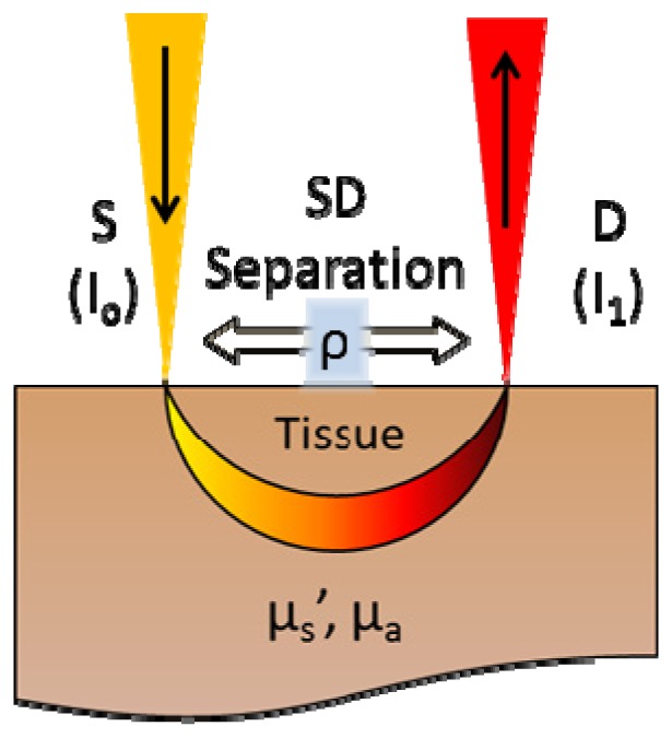
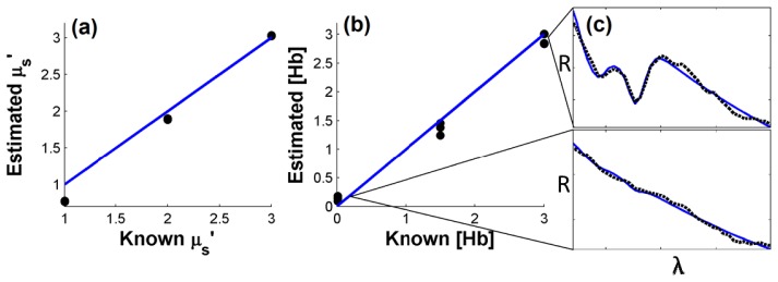
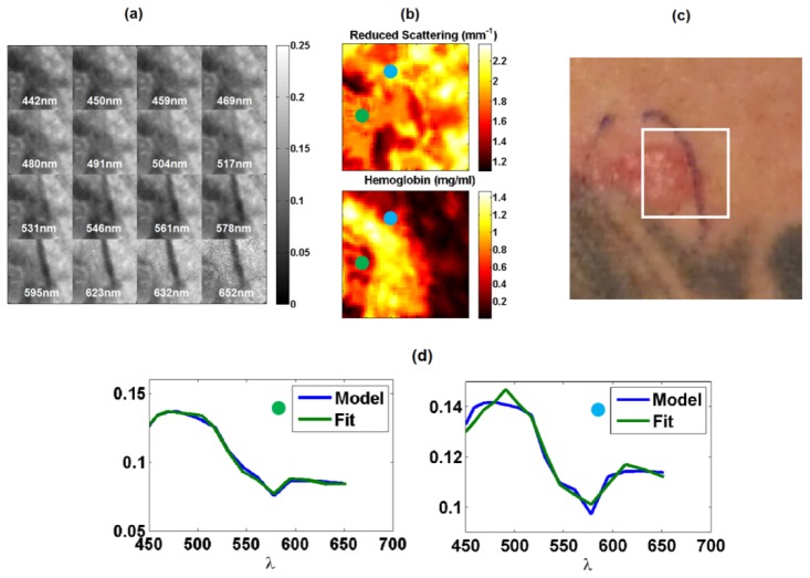
Diffuse reflectance spectroscopy provides a noninvasive means to measure optical and physiological properties of tissues. To expand on these measurements, we have developed a handheld diffuse reflectance spectral imaging (DRSi) system capable of acquiring wide field hyperspectral images of tissue. The image acquisition time was approximately 50 seconds for a 50x50 pixel image. A transport model was used to fit each spectra for reduced scattering coefficient, hemoglobin concentration and melanin concentration resulting in optical property maps. The system was validated across biologically relevant levels of reduced scattering (5.14% error) and absorption (8.34% error) using tissue simulating phantoms. DRSi optical property maps of a pigmented skin lesion were acquired in vivo. These trends in optical properties were consistent with previous observations using point probe devices.
Keywords: (110.0110) Imaging systems; (110.0113) Imaging through turbid media; (170.6510) Spectroscopy, tissue diagnostics.
Figures


References
-
- Martin M. E., Wabuyele M. B., Chen K., Kasili P., Panjehpour M., Phan M., Overholt B., Cunningham G., Wilson D., Denovo R. C., Vo-Dinh T., “Development of an advanced hyperspectral imaging (HSI) system with applications for cancer detection,” Ann. Biomed. Eng. 34(6), 1061–1068 (2006).10.1007/s10439-006-9121-9 - DOI - PubMed
-
- D. Roblyer, C. Kurachi, A. M. Gillenwater, and R. Richards-Kortum, “In Vivo Fluorescence Hyperspectral Imaging of Oral Neoplasia,” in Advanced Biomedical and Clinical Diagnostic Systems Vii, A. MahadevanJansen, T. VoDinh, and W. S. Grundfest, eds. (2009).
Grants and funding
LinkOut - more resources
Full Text Sources
Other Literature Sources