

doi: 10.1155/2014/920538.
Epub 2014 Jan 19.
A standardized method for 4D ultrasound-guided peripheral nerve blockade and catheter placement
Affiliations
- PMID: 24575416
- PMCID: PMC3915798
- DOI: 10.1155/2014/920538
Item in Clipboard
A standardized method for 4D ultrasound-guided peripheral nerve blockade and catheter placement
Biomed Res Int.
2014.
Abstract
We present a standardized method for using four-dimensional ultrasound (4D US) guidance for peripheral nerve blocks. 4D US allows for needle tracking in multiple planes simultaneously and accurate measurement of the local anesthetic volume surrounding the nerve following injection. Additionally, the morphology and proximity of local anesthetic spread around the target nerve is clearly seen with the described technique. This method provides additional spatial information in real time compared to standard two-dimensional ultrasound.
Figures
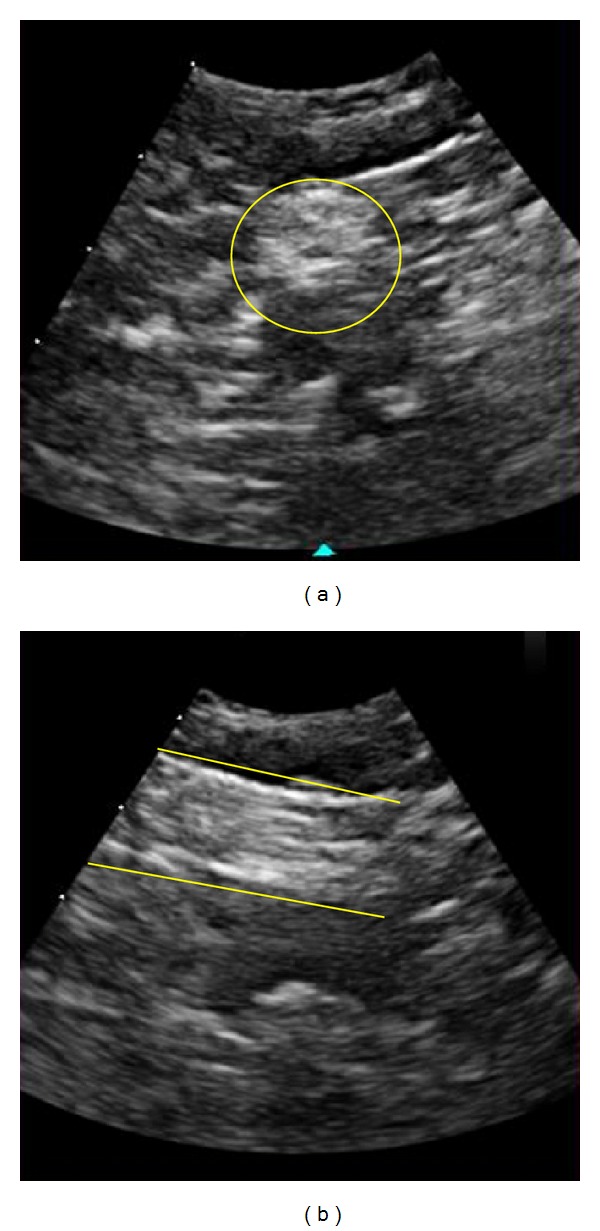
Image of the sciatic nerve at the popliteal fossa in short and long axis view obtained simultaneously in x-Plane mode. The yellow outlines are added for clarity.
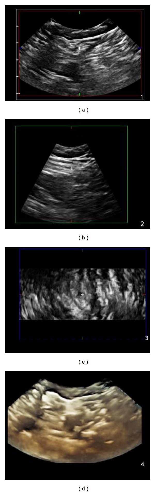
Standard view of the sciatic nerve from the popliteal fossa in 4D mode. The white crosshair corresponds to the same location on the sciatic nerve in short axis (a), long axis (b), and coronal view (c). (d) is a three-dimensional composite image that updates in real time.
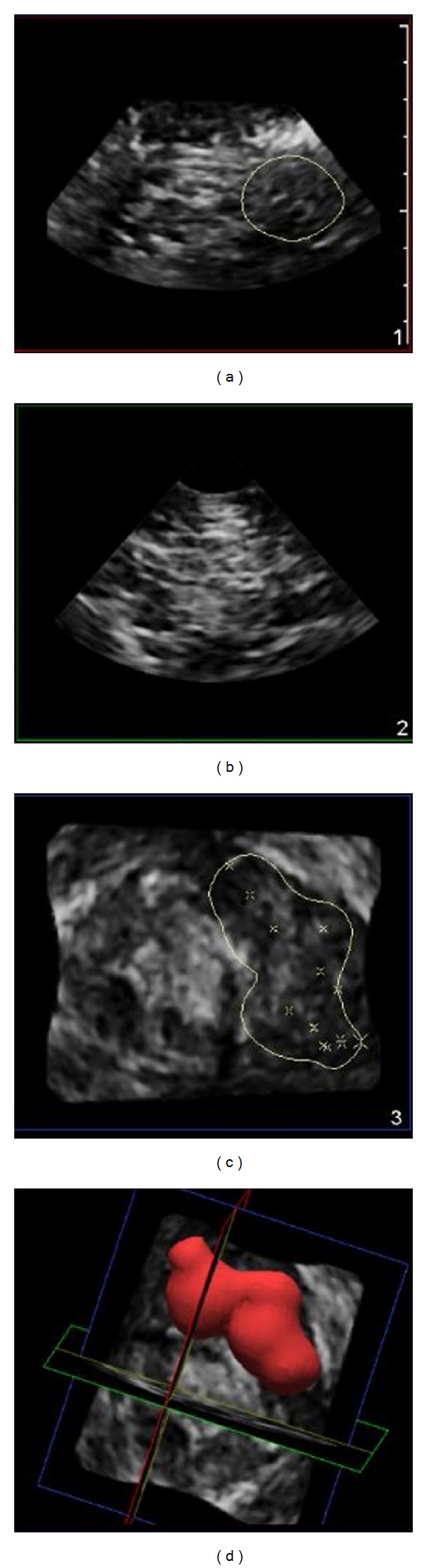
Infraclavicular brachial plexus block volume calculated as 21.2 milliliters demonstrating epineural local anesthetic spread. Injected local anesthetic volume was 20 milliliters.
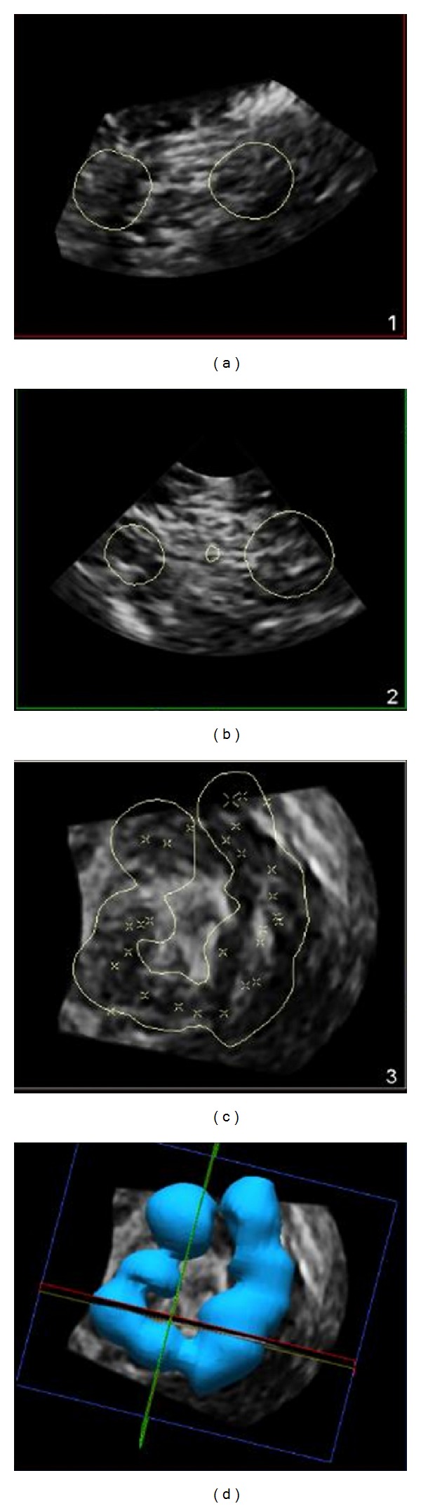
Infraclavicular brachial plexus block volume calculated as 41 milliliters demonstrating circumneural local anesthetic spread. Injected local anesthetic volume was 40 milliliters.
Similar articles
-
A prospective, randomized comparison between single- and multiple-injection techniques for ultrasound-guided subgluteal sciatic nerve block.Anesth Analg. 2014 Dec;119(6):1442-8. doi: 10.1213/ANE.0000000000000462. Anesth Analg. 2014. PMID: 25268398 Clinical Trial.
-
Ultrasound-guided peripheral nerve blockade.Curr Pain Headache Rep. 2007 Feb;11(1):25-32. doi: 10.1007/s11916-007-0018-6. Curr Pain Headache Rep. 2007. PMID: 17214918 Review.
-
Case report: Three-dimensional high-resolution ultrasound-guided nerve blocks: a new panoramic vision of local anesthetic spread and perineural catheter tip location.Anesth Analg. 2013 May;116(5):1176-1181. doi: 10.1213/ANE.0b013e31828b34ae. Epub 2013 Mar 14. Anesth Analg. 2013. PMID: 23492963
-
Electrical stimulation versus ultrasound guidance for popliteal-sciatic perineural catheter insertion: a randomized controlled trial.Reg Anesth Pain Med. 2009 Sep-Oct;34(5):480-5. doi: 10.1097/AAP.0b013e3181ada57a. Reg Anesth Pain Med. 2009. PMID: 19920423 Clinical Trial.
-
Ultrasonography and stimulating perineural catheters for nerve blocks: a review of the evidence.Can J Anaesth. 2008 Jul;55(7):447-57. doi: 10.1007/BF03016312. Can J Anaesth. 2008. PMID: 18591703 Review.
Cited by
-
"Knowing It Before Blocking It," the ABCD of the Peripheral Nerves: Part C (Prevention of Nerve Injuries).Cureus. 2023 Jul 13;15(7):e41847. doi: 10.7759/cureus.41847. eCollection 2023 Jul. Cureus. 2023. PMID: 37581128 Free PMC article. Review.
-
Thyroid gland visualization with 3D/4D ultrasound: integrated hands-on imaging in anatomical dissection laboratory.Surg Radiol Anat. 2017 May;39(5):567-572. doi: 10.1007/s00276-016-1775-x. Epub 2016 Dec 1. Surg Radiol Anat. 2017. PMID: 27909799 Free PMC article.
-
Assessment in location of sciatic nerve between the ischial tuberosity and the greater trochanter of the femur: A cadaveric study.Heliyon. 2023 Dec 16;10(1):e23751. doi: 10.1016/j.heliyon.2023.e23751. eCollection 2024 Jan 15. Heliyon. 2023. PMID: 38192877 Free PMC article.
-
Development of technologies for placement of perineural catheters.J Anesth. 2016 Feb;30(1):138-47. doi: 10.1007/s00540-015-2076-y. Epub 2015 Sep 14. J Anesth. 2016. PMID: 26370264 Review.
References
-
- Choquet O, Capdevila X. Case report: three-dimensional high-resolution ultrasound-guided nerve blocks: a new panoramic vision of local anesthetic spread and perineural catheter tip location. Anesthesia & Analgesia. 2013;116(5):1176–1181. - PubMed
-
- Albrecht H, Stroszczynski C, Felix R, Hünerbein M. Real time 3D (4D) ultrasound-guided percutaneous biopsy of solid tumours. Ultraschall in der Medizin. 2006;27(4):324–328. - PubMed
-
- Sugimoto K, Moriyasu F, Shiraishi J, Yamada M, Imai Y. A phantom study comparing ultrasound-guided liver tumor puncture using new real-time 3D ultrasound and conventional 2D ultrasound. American Journal of Roentgenology. 2011;196(6):W753–W757. - PubMed
-
- Clendenen SR, Riutort K, Ladlie BL, Robards C, Franco CD, Greengrass RA. Real-time three-dimensional ultrasound-assisted axillary plexus block defines soft tissue planes. Anesthesia & Analgesia. 2009;108(4):1347–1350. - PubMed
-
- Feinglass NG, Clendenen SR, Torp KD, Wang RD, Castello R, Greengrass RA. Real-time three-dimensional ultrasound for continuous popliteal blockade: a case report and image description. Anesthesia & Analgesia. 2007;105(1):272–274. - PubMed
MeSH terms
LinkOut - more resources
Full Text Sources
Other Literature Sources
