

Strength of axial water ligation in substrate-free cytochrome P450s is isoform dependent
- PMID: 24576089
- PMCID: PMC3985942
- DOI: 10.1021/bi401547j
Strength of axial water ligation in substrate-free cytochrome P450s is isoform dependent
Abstract
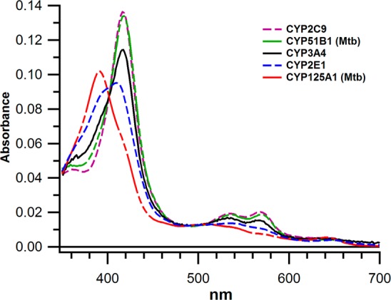
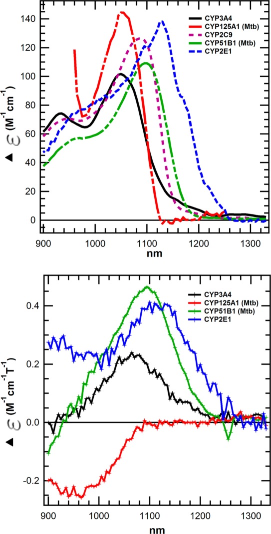
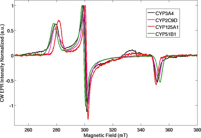
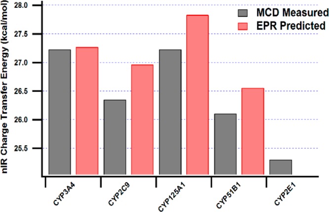
The heme-containing cytochrome P450s exhibit isoform-dependent ferric spin equilibria in the resting state and differential substrate-dependent spin equilibria. The basis for these differences is not well understood. Here, magnetic circular dichroism (MCD) reveals significant differences in the resting low spin ligand field of CYPs 3A4, 2E1, 2C9, 125A1, and 51B1, which indicates differences in the strength of axial water ligation to the heme. The near-infrared bands that specifically correspond to charge-transfer porphyrin-to-metal transitions span a range of energies of nearly 2 kcal/mol. In addition, the experimentally determined MCD bands are not entirely in agreement with the expected MCD energies calculated from electron paramagnetic resonance parameters, thus emphasizing the need for the experimental data. MCD marker bands of the high spin heme between 500 and 680 nm were also measured and suggest only a narrow range of energies for this ensemble of high spin Cys(S(-)) → Fe(3+) transitions among these isoforms. The differences in axial ligand energies between CYP isoforms of the low spin states likely contribute to the energetics of substrate-dependent spin state perturbation. However, these ligand field energies do not correlate with the fraction of high spin vs low spin in the resting state enzyme, suggestive of differences in water access to the heme or isoform-dependent differences in the substrate-free high spin states as well.
Figures


References
-
- Sono M.; Roach M. P.; Coulter E. D.; Dawson J. H. (1996) Heme-containing oxygenases. Chem. Rev. 96, 2841–2888. - PubMed
-
- Guengerich F. P. (2001) Common and uncommon cytochrome P450 reactions related to metabolism and chemical toxicity. Chem. Res. Toxicol. 14, 611–650. - PubMed
-
- Sligar S. G. (1976) Coupling of spin, substrate, and redox equilibriums in cytochrome P450. Biochemistry 15, 5399–5406. - PubMed
-
- Fisher M. T.; Sligar S. G. (1985) Control of heme protein redox potential and reduction rate: linear free energy relation between potential and ferric spin state equilibrium. J. Am. Chem. Soc. 107, 5018–5019.
-
- Roberts A. G.; Campbell A. P.; Atkins W. M. (2005) The thermodynamic landscape of testosterone binding to cytochrome P450 3A4: ligand binding and spin state equilibria. Biochemistry 44, 1353–1366. - PubMed
Publication types
MeSH terms
Substances
Grants and funding
LinkOut - more resources
Full Text Sources
Other Literature Sources
