Review
. 2014 Mar 15;458(3):421-37.
doi: 10.1042/BJ20140006.

RBR E3 ubiquitin ligases: new structures, new insights, new questions

Affiliations
Review

RBR E3 ubiquitin ligases: new structures, new insights, new questions

Donald E Spratt et al. Biochem J. .

Abstract

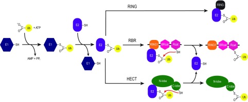
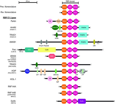
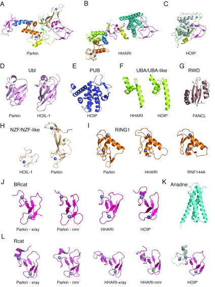
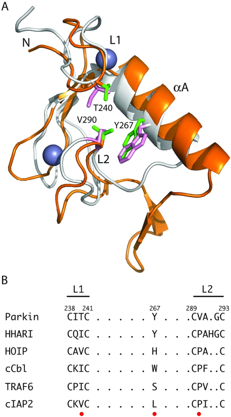
The RBR (RING-BetweenRING-RING) or TRIAD [two RING fingers and a DRIL (double RING finger linked)] E3 ubiquitin ligases comprise a group of 12 complex multidomain enzymes. This unique family of E3 ligases includes parkin, whose dysfunction is linked to the pathogenesis of early-onset Parkinson's disease, and HOIP (HOIL-1-interacting protein) and HOIL-1 (haem-oxidized IRP2 ubiquitin ligase 1), members of the LUBAC (linear ubiquitin chain assembly complex). The RBR E3 ligases share common features with both the larger RING and HECT (homologous with E6-associated protein C-terminus) E3 ligase families, directly catalysing ubiquitin transfer from an intrinsic catalytic cysteine housed in the C-terminal domain, as well as recruiting thioester-bound E2 enzymes via a RING domain. Recent three-dimensional structures and biochemical findings of the RBRs have revealed novel protein domain folds not previously envisioned and some surprising modes of regulation that have raised many questions. This has required renaming two of the domains in the RBR E3 ligases to more accurately reflect their structures and functions: the C-terminal Rcat (required-for-catalysis) domain, essential for catalytic activity, and a central BRcat (benign-catalytic) domain that adopts the same fold as the Rcat, but lacks a catalytic cysteine residue and ubiquitination activity. The present review discusses how three-dimensional structures of RBR (RING1-BRcat-Rcat) E3 ligases have provided new insights into our understanding of the biochemical mechanisms of these important enzymes in ubiquitin biology.


Figures

Figure 1
Figure 1. Proposed pathways for ubiquitination by different E3 ubiquitin ligases
The ubiquitin activating enzyme (E1) activates ubiquitin through an ATP-dependent mechanism to form a thioester bond between the C-terminal carboxyl of ubiquitin and the catalytic cysteine in the E1. The ubiquitin is then transferred to an ubiquitin conjugating enzyme (E2) via a transthiolation reaction to form a thioester bond between the C-terminus of ubiquitin and the conserved catalytic cysteine residue of the E2. For the RING E3 ligases, the E2~ubiquitin complex engages with the RING domain of the E3 which optimally positions the ubiquitin in preparation for its transfer to a substrate protein. The HECT E3 ligases engage the E2~ubiquitin complex via their N-terminal lobe and perform another transthiolation reaction to form a thioester bond between the C-terminus of ubiquitin and the conserved catalytic cysteine residue in the C-terminal lobe of the HECT E3s. This HECT~ubiquitin intermediate is then poised for the subsequent transfer of ubiquitin to a substrate. The RBR E3 ligases use a combination of the RING and HECT mechanisms (termed a ‘RING–HECT’ hybrid mechanism [29]). In this mechanism, the RING1 engages with the E2~ubiquitin complex in a similar manner to the RING E3s, whereas the Rcat acts in a similar fashion to the C-terminal lobe of the HECT E3s by performing a transthiolation reaction to form a thiolester bond between the C-terminus of ubiquitin and the catalytic cysteine of the Rcat domain of RBR E3s.
Figure 2
Figure 2. Domain architecture of the RBR E3 ubiquitin ligases
Domains found in each RBR E3 ligase are RING1 (orange) BRcat (previously known as IBR; hot pink), and Rcat, (previously known as RING2; warm pink). Other domains listed include the Ubl (light pink), RING0 (wheat) and Npl4 NZF (wheat), acidic/Gly N-terminal extension (Acidic/Gly or Acidic; dark green), UBA-like (lime green), Ariadne domain (cyan), UIM (olive), ankyrin repeats (dark grey), docking domain (DOC; teal), Cullin (pale yellow), RWD (dark salmon), PUB (deep blue), ZnF (pale green), helical base (light grey) and Dorfin domain (deep purple). A conserved domain found in Cul7, Parc, and HERC2 proteins (CPH) is located in the N-terminal extension of Parc (not shown).
Figure 3
Figure 3. Catalogue of three-dimensional structures for RBR E3 ubiquitin ligases
The upper panels show cartoon representations of multi-domain structures for (A) RING0–RBR from human parkin (PDB code 4I1F [35]; also PDB code 4K7D [34] and PDB code 4BM9 [36]), (B) human HHARI (PDB code 4KBL [42]) and (C) C-terminus of human HOIP (PDB code 4LJP [43]). The lower panels (DL) show cartoon diagrams of three-dimensional structures of the individual domains for (D) Ubl domains from parkin (PDB code 2ZEQ [136]) and HOIL-1 (PDB code 2LGY [81]), (E) PUB domain from HOIP (PDB code 4JUY), (F) UBA-like domains from HHARI (PDB code 4KBL [42]) and HOIP (PDB code 4DBG [32]), (G) RWD from the E3 ligase FANCL (PDB code 3K1L [108]), (H) NZF and double NZF-like domains from HOIL-1 (PDB code 3B0A [39]) and parkin (PDB code 4I1F [35]), (I) RING1 domains from parkin (PDB code 4I1F [35]), HHARI (PDB code 4KBL [42]) and RNF144A (PDB code 1WIM), (J) BRcat domains from parkin (PDB code 4I1F [35] and PDB 2JMO [116]), HHARI (PDB code 4KBL [42]) and HOIP (PDB code 2CT7), (K) Ariadne domain from HHARI (PDB code 4KBL [42]), and (L) Rcat domains from parkin (PDB code 4I1F [35] and PDB code 2LWR [48]), HHARI (PDB code 4KBL [42] and PDB 2M9Y [117]) and HOIP (PDB code 4LJP [43]). The colour scheme for each individual domain and multidomain structures are as shown in Figure 2. Representative secondary structures are also labelled.
Figure 4
Figure 4. Comparison of RING domain structures for RBR and canonical RING E3 ubiquitin ligases
(A) The structures of the RING1 domain from parkin (PDB code 4I1F [35]; orange) is superimposed with the RING domain from c-Cbl (PDB code 1FBV [5]; grey). The superposition was done using the Cα positions of the eight Zn2+-co-ordinating residues in each protein. The two regions (L1 and L2) in each protein and residues in parkin expected to be key for E2 interaction are indicated. (B) Sequence comparison for the RING1 domains of the RBR proteins parkin, HHARI and HOIP with representative RING E3 ligases c-Cbl, TRAF6 and cIAP2 showing important residues for E2 recruitment in L1 and L2 loops (red dot).
Figure 5
Figure 5. Similarity of catalytic sites for parkin and NEDD4
(A) The interface between the Rcat (pink ribbon) and RING0 (wheat surface) domains for parkin are shown highlighting important residues near the catalytic site. The three residues (Cys431, His433 and Glu444) important for ubiquitin transfer are shown in addition to several residues found at the Rcat (Trp462 and Phe463), RING0 (Lys162, Trp183, Pro180 and Val186) interface (PDB code 4I1F [35]). (B) A portion of the interface between the N-lobe (grey surface) and the catalytic region of the C-lobe (green ribbon) in NEDD4 is shown (PDB code 4BBN [122]). The catalytic cysteine (Cys867) resides between two β-strands similar to the position in parkin and HHARI. Two other residues important for catalysis (His865 and Asp900) are arranged in a mirror fashion compared to the Rcat domain in parkin and HHARI although Asp900 is not visible in the X-ray structure. In both structures the two β-strands were superimposed to achieve similar protein orientations.
Figure 6
Figure 6. Evidence for flexibility in the RBR E3 ubiquitin ligases
(A) The three-dimensional structures of parkin (PDB code 4I1F [35]) and HHARI (PDB code 4KBL [42]) are shown following superposition of their RING1 domains. This presentation shows the Rcat domains for the two proteins are found at opposite ends of the respective structures with respect to the location of the RING1 domain with large distances between the RING1 and Rcat domains (~32 Å in parkin and ~67 Å in HHARI) that must somehow be bridged for ubiquitin transfer. Regions not modelled in the parkin structure, probably due to flexibility, are indicated by broken lines. For clarity the UBA-like and Ariadne domains from HHARI are not shown. (B) The position of the BRcat domains in parkin and HHARI are shown after superposition of the RING1 domains. The position of the BRcat domain deviates ~22 Å between the parkin and HHARI structures due to an approximate 90° difference in the tilt of the RING1–BRcat interdomain helix. Only the RING1 domain from parkin is shown for clarity. The colour schemes used in (A) and (B) are as described previously in Figures 2 and 3.

References

    1. Hershko A., Ciechanover A. The ubiquitin system. Annu. Rev. Biochem. 1998;67:425–479. - PubMed
    1. Deshaies R. J., Joazeiro C. A. RING domain E3 ubiquitin ligases. Annu. Rev. Biochem. 2009;78:399–434. - PubMed
    1. Metzger M. B., Pruneda J. N., Klevit R. E., Weissman A. M. RING-type E3 ligases: master manipulators of E2 ubiquitin-conjugating enzymes and ubiquitination. Biochim. Biophys. Acta. 2014;1843:47–60. - PMC - PubMed
    1. Borden K. L., Freemont P. S. The RING finger domain: a recent example of a sequence-structure family. Curr. Opin. Struct. Biol. 1996;6:395–401. - PubMed
    1. Zheng N., Wang P., Jeffrey P. D., Pavletich N. P. Structure of a c-Cbl–UbcH7 complex: RING domain function in ubiquitin-protein ligases. Cell. 2000;102:533–539. - PubMed

Publication types