. 2014 Feb 28:13:41.
doi: 10.1186/1476-4598-13-41.

The taccalonolides and paclitaxel cause distinct effects on microtubule dynamics and aster formation

Affiliations

The taccalonolides and paclitaxel cause distinct effects on microtubule dynamics and aster formation

April L Risinger et al. Mol Cancer. .

Abstract

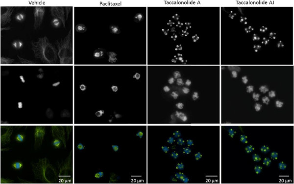
Background: Microtubule stabilizers suppress microtubule dynamics and, at the lowest antiproliferative concentrations, disrupt the function of mitotic spindles, leading to mitotic arrest and apoptosis. At slightly higher concentrations, these agents cause the formation of multiple mitotic asters with distinct morphologies elicited by different microtubule stabilizers.

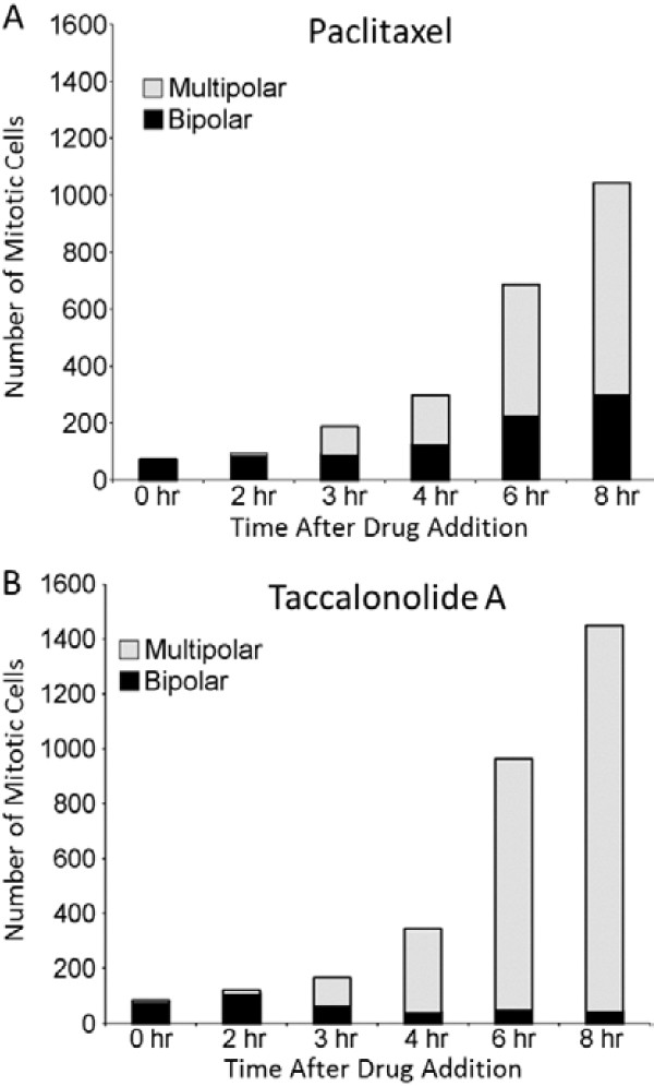
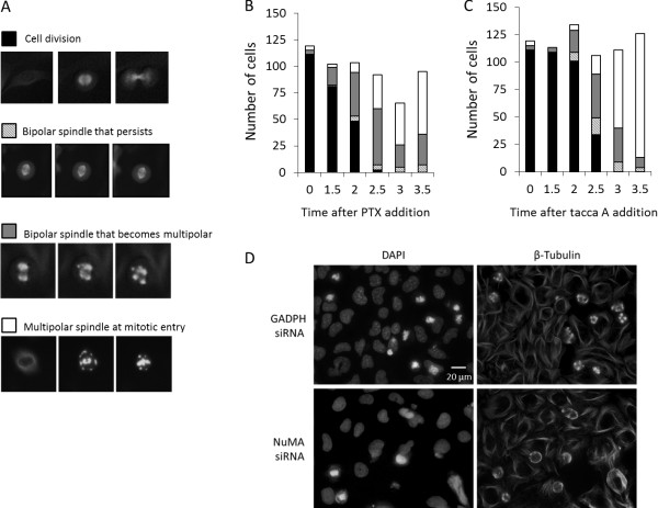
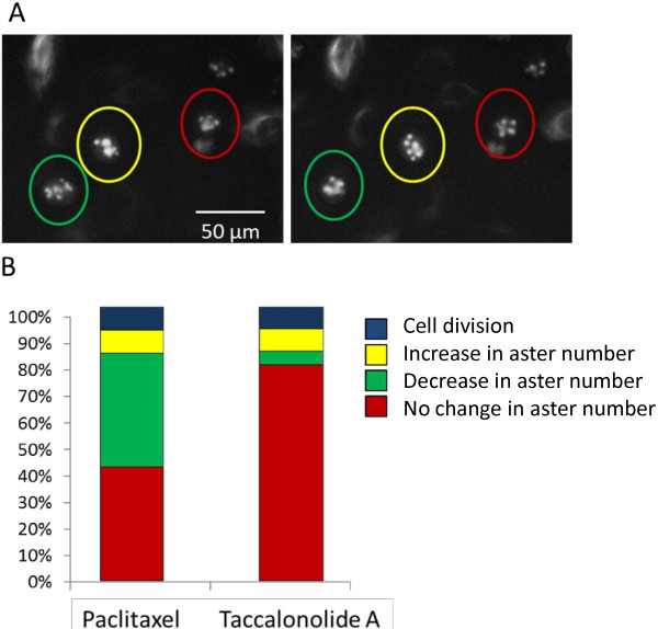
Results: We tested the hypothesis that two classes of microtubule stabilizing drugs, the taxanes and the taccalonolides, cause the formation of distinct aster structures due, in part, to differential effects on microtubule dynamics. Paclitaxel and the taccalonolides suppressed the dynamics of microtubules formed from purified tubulin as well as in live cells. Both agents suppressed microtubule dynamic instability, with the taccalonolides having a more pronounced inhibition of microtubule catastrophe, suggesting that they stabilize the plus ends of microtubules more effectively than paclitaxel. Live cell microscopy was also used to evaluate the formation and resolution of asters after drug treatment. While each drug had similar effects on initial formation, substantial differences were observed in aster resolution. Paclitaxel-induced asters often coalesced over time resulting in fewer, larger asters whereas numerous compact asters persisted once they were formed in the presence of the taccalonolides.

Conclusions: We conclude that the increased resistance of microtubule plus ends to catastrophe may play a role in the observed inability of taccalonolide-induced asters to coalesce during mitosis, giving rise to the distinct morphologies observed after exposure to these agents.


Figures

Figure 1
Figure 1
Mitotic asters induced by paclitaxel or the taccalonolides. The mitotic asters in HeLa cells treated with vehicle, 12 nM paclitaxel, 5 μM taccalonolide A or 20 nM taccalonolide AJ, the minimum concentrations that caused maximum mitotic accumulation, were visualized by indirect immunofluorescence for β-tubulin (top). DNA was visualized by DAPI staining (middle) and merged images are shown with microtubules in green and DNA in blue (bottom).
Figure 2
Figure 2
Effects of paclitaxel or taccalonolide A on mitotic accumulation and aster number. The number of mitotic cells present at each time after the addition of (A) 12 nM paclitaxel or (B) 5 μM taccalonolide A to GFP-β-tubulin expressing HeLa cells was counted. These mitotic cells were classified as containing bipolar spindles (black) or multiple asters (gray). Cells from 60 microscopic fields (20x) from 12 individual wells were analyzed for each condition at each time point.
Figure 3
Figure 3
Aster formation following the addition of paclitaxel or taccalonolide A. GFP-β-tubulin expressing HeLa cells were treated with 12 nM paclitaxel or 5 μM taccalonolide A. Every cell that entered mitosis within 3.5 h after drug addition was followed until 8 h after drug addition and placed in one of 4 categories. (A) Representative images of the 4 categories of cells entering mitosis: bipolar spindle formation followed by cell division, bipolar spindle formation that persisted without completion of mitosis, bipolar spindle that resolved into multiple asters or formation of multiple asters immediately upon mitotic entry. Each image represents approximately 40 μm. The category of cells entering mitosis at the indicated time points following (B) paclitaxel or (C) taccalonolide A addition are shown. Cells in 60 individual microscopic fields from 12 separate wells were analyzed for each condition at each time point. (D) Effects of NuMA depletion on taccalonolide A-induced asters. Microtubules were visualized by indirect immunofluorescence 4 h after the addition of 5 μM taccalonolide A in control (GADPH siRNA) and NuMA-depleted (NuMA siRNA) HeLa cells (right). DNA was visualized by DAPI staining (left).
Figure 4
Figure 4
Resolution of drug induced asters. The resolution of the multiple asters formed in GFP-β-tubulin expressing HeLa cells after the addition of 12 nM paclitaxel or 5 μM taccalonolide A were evaluated 5–8 h after drug addition. (A) A representative microscope field showing mitotic cells that undergo: 1) a decrease in aster number (green circle), 2) increase in aster number (yellow circle) or 3) no change in number of asters (red circle) are indicated. (B) Percentage of mitotic cells that undergo each phenotype or successfully divide during the time period. 100 cells were followed over time for each condition.

References

    1. Ikui AE, Yang CP, Matsumoto T, Horwitz SB. Low concentrations of taxol cause mitotic delay followed by premature dissociation of p55CDC from Mad2 and BubR1 and abrogation of the spindle checkpoint, leading to aneuploidy. Cell Cycle. 2005;4:1385–1388. doi: 10.4161/cc.4.10.2061. - DOI - PubMed
    1. Yang Z, Kenny AE, Brito DA, Rieder CL. Cells satisfy the mitotic checkpoint in Taxol, and do so faster in concentrations that stabilize syntelic attachments. J Cell Biol. 2009;186:675–684. doi: 10.1083/jcb.200906150. - DOI - PMC - PubMed
    1. Gascoigne KE, Taylor SS. Cancer cells display profound intra- and interline variation following prolonged exposure to antimitotic drugs. Cancer Cell. 2008;14:111–122. doi: 10.1016/j.ccr.2008.07.002. - DOI - PubMed
    1. Risinger AL, Mooberry SL. Cellular studies reveal mechanistic differences between taccalonolide A and paclitaxel. Cell Cycle. 2011;10:2162–2171. doi: 10.4161/cc.10.13.16238. - DOI - PMC - PubMed
    1. Schiff PB, Horwitz SB. Taxol stabilizes microtubules in mouse fibroblast cells. Proc Natl Acad Sci U S A. 1980;77:1561–1565. doi: 10.1073/pnas.77.3.1561. - DOI - PMC - PubMed

Publication types

LinkOut - more resources