Review
. 2014 Mar 18;53(10):1565-74.
doi: 10.1021/bi5000564. Epub 2014 Mar 7.

Mechanism of quinolone action and resistance

Affiliations
Review

Mechanism of quinolone action and resistance

Katie J Aldred et al. Biochemistry. .

Abstract

Quinolones are one of the most commonly prescribed classes of antibacterials in the world and are used to treat a variety of bacterial infections in humans. Because of the wide use (and overuse) of these drugs, the number of quinolone-resistant bacterial strains has been growing steadily since the 1990s. As is the case with other antibacterial agents, the rise in quinolone resistance threatens the clinical utility of this important drug class. Quinolones act by converting their targets, gyrase and topoisomerase IV, into toxic enzymes that fragment the bacterial chromosome. This review describes the development of the quinolones as antibacterials, the structure and function of gyrase and topoisomerase IV, and the mechanistic basis for quinolone action against their enzyme targets. It will then discuss the following three mechanisms that decrease the sensitivity of bacterial cells to quinolones. Target-mediated resistance is the most common and clinically significant form of resistance. It is caused by specific mutations in gyrase and topoisomerase IV that weaken interactions between quinolones and these enzymes. Plasmid-mediated resistance results from extrachromosomal elements that encode proteins that disrupt quinolone-enzyme interactions, alter drug metabolism, or increase quinolone efflux. Chromosome-mediated resistance results from the underexpression of porins or the overexpression of cellular efflux pumps, both of which decrease cellular concentrations of quinolones. Finally, this review will discuss recent advancements in our understanding of how quinolones interact with gyrase and topoisomerase IV and how mutations in these enzymes cause resistance. These last findings suggest approaches to designing new drugs that display improved activity against resistant strains.


Figures

Figure 1
Figure 1
Quinolone structures. Nalidixic acid and oxolinic acid were the first-generation quinolones that were used most often in the clinic. Norfloxacin, ciprofloxacin, and ofloxacin are the most relevant second-generation quinolones. Levofloxacin (the levorotary isomer of ofloxacin), sparfloxacin, and moxifloxacin are newer-generation quinolones.
Figure 2
Figure 2
Domain structures of type II topoisomerases. Gyrase and topoisomerase IV are heterotetrameric enzymes consisting of two A subunits and two B subunits. The A subunits (blue and red; GyrA in gyrase and ParC and GrlA in Gram-negative and Gram-positive topoisomerase IV, respectively) contain the active site tyrosine residue that covalently attaches to the newly generated 5′-termini of DNA during the cleavage reaction. The C-terminal domains (CTDs; red) of the A subunits are variable and allow gyrase to introduce negative supercoils into DNA. The B subunits (green; GyrB in gyrase and ParE and GrlB in Gram-negative and Gram-positive topoisomerase IV, respectively) contain the ATPase and TOPRIM domains, the latter of which binds the catalytic divalent metal ions essential for enzyme activity. Human topoisomerase IIα is homologous to the bacterial type II enzymes. However, during the course of evolution, the A and B subunits fused into a single polypeptide chain. Therefore, eukaryotic type II topoisomerases function as homodimers. A representation of the three-dimensional structure of type II topoisomerases is shown at the bottom.
Figure 3
Figure 3
Crystal structure of a moxifloxacin-stabilized Acinetobacter baumannii topoisomerase IV–DNA cleavage complex. The catalytic core of the enzyme is shown. Moxifloxacin is colored red; the topoisomerase IV A and B subunits are colored blue and green, respectively, and DNA is colored yellow. The top panel is a top view of the cleavage complex showing two quinolone molecules intercalating 4 bp apart at the sites of DNA cleavage. The bottom panel is a front view (rotated by 90° from the top view) of the cleavage complex. Protein Data Bank accession 2XKK was visualized using Discovery Studio 3.5 Visualizer (Accelrys Software Inc.). Adapted from ref (11).
Figure 4
Figure 4
Bacterial type II topoisomerases are essential but potentially toxic enzymes. The balance between enzyme-mediated DNA cleavage and religation is critical for cell survival. If the level of gyrase-mediated DNA cleavage decreases, rates of DNA replication slow and impair cell growth (left). If the level of topoisomerase IV-mediated DNA cleavage decreases, cells are not able to untangle daughter chromosomes and ultimately die of mitotic failure (left). If the level of gyrase- or topoisomerase IV-mediated DNA cleavage becomes too high (right), the actions of DNA tracking systems can convert these transient complexes to permanent double-stranded breaks. The resulting DNA breaks initiate the SOS response and other DNA repair pathways and can lead to cell death.
Figure 5
Figure 5
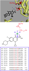
Quinolone–topoisomerase binding is facilitated through a water–metal ion bridge. The top panel shows the crystal structure of a moxifloxacin-stabilized A. baumannii topoisomerase IV–DNA cleavage complex. Moxifloxacin is colored black, and the noncatalytic Mg2+ ion that is chelated by the C3/C4 keto acid of the quinolone and participates in the bridge interaction is colored green. The four water molecules that fill out the coordination sphere of the Mg2+ ion are colored blue. The backbone of selected portions of the protein amino acid chain is colored yellow. The side chains of the serine and acidic residues that form hydrogen bonds with the water molecules in the water–metal ion bridge are colored red. In A. baumannii topoisomerase IV, these residues are Ser84 and Glu88, respectively. For clarity, DNA has been omitted from the picture. Protein Data Bank accession 2XKK was visualized using Discovery Studio 3.5 Visualizer (Accelrys Software Inc.). Adapted from ref (11). The middle panel shows a simplified diagram of the water–metal ion bridge. Only interactions with the protein (and not DNA) are shown. Ciprofloxacin (a representative quinolone) is colored black, and the noncatalytic Mg2+, water molecules, and coordinating serine and acidic residues are colored as described above. Blue dashed lines indicate the octahedral coordination sphere of the divalent metal ion interacting with four water molecules and the C3/C4 keto acid of the quinolone. The red dashed lines represent hydrogen bonds between the serine side chain hydroxyl group or the acidic residue side chain carboxyl group and the water molecules. Adapted from ref (13). The bottom panel shows a sequence alignment of the A subunits highlighting the conserved serine and acidic residues (red) that coordinate the water–metal ion bridge. Sequences of A. baumannii (Ab), Bacillus anthracis (Ba), E. coli (Ec), Staphylococcus aureus (Sa), and S. pneumoniae (Sp) gyrase (GyrA) and topoisomerase IV (ParC/GrlA) are shown. The homologous regions of human topoisomerase IIα (hTIIα) and IIβ (hTIIβ), which lack the residues necessary to coordinate the water–metal ion bridge interaction, are shown for comparison.
Figure 6
Figure 6
Mechanisms of quinolone resistance. (1) Target-mediated resistance. Mutations in gyrase and topoisomerase IV weaken quinolone–enzyme interactions. (2) Plasmid-mediated resistance. (2a) Qnr proteins (yellow) decrease topoisomerase–DNA binding and protect enzyme–DNA complexes from quinolones. (2b) Aac(6′)-Ib-cr is an aminoglycoside acetyltransferase that acetylates the free nitrogen on the C7 ring of ciprofloxacin and norfloxacin, decreasing their effectiveness. (2c) Plasmid-encoded efflux pumps decrease the concentration of quinolones in the cell. (3) Chromosome-mediated resistance. (3a) Underexpression of porins in Gram-negative species decreases drug uptake. (3b) Overexpression of chromosome-encoded efflux pumps decreases drug retention in the cell.
Figure 7
Figure 7
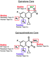
Roles of substituents and core elements of quinolones and quinazolinediones that mediate drug activity against bacterial and human type II topoisomerases. Results are based on studies with B. anthracis topoisomerase IV and human topoisomerase IIα. For quinolones, the binding of clinically relevant drugs to topoisomerase IV is mediated primarily through the water–metal ion bridge. The binding of quinolones that overcome resistance is mediated primarily by the substituent at C7. Binding of quinolones to human topoisomerase IIα also is mediated by the C7 substituent. The group at C8 affects the ability of quinolones to act against the human type II enzyme but is not required for drug binding. For quinazolinediones, interactions between drugs and topoisomerase IV (wild-type and resistant) are mediated through the C7 substituent. The effects of the C7 and C8 substituents on quinazolinedione activity against topoisomerase IIα are the same as described for the quinolones. The N3 amino group plays a role in the binding of quinazolinediones to the human enzyme. Adapted from ref (53).

References

    1. Emmerson A. M.; Jones A. M. (2003) The quinolones: Decades of development and use. J. Antimicrob. Chemother. 51(Suppl. 1), 13–20. - PubMed
    1. Mitscher L. A. (2005) Bacterial topoisomerase inhibitors: Quinolone and pyridone antibacterial agents. Chem. Rev. 105, 559–592. - PubMed
    1. Linder J. A.; Huang E. S.; Steinman M. A.; Gonzales R.; Stafford R. S. (2005) Fluoroquinolone prescribing in the United States: 1995 to 2002. Am. J. Med. 118, 259–268. - PubMed
    1. Andriole V. T. (2005) The quinolones: Past, present, and future. Clin. Infect. Dis. 41(Suppl. 2), S113–S119. - PubMed
    1. Drlica K.; Hiasa H.; Kerns R.; Malik M.; Mustaev A.; Zhao X. (2009) Quinolones: Action and resistance updated. Curr. Top. Med. Chem. 9, 981–998. - PMC - PubMed

Publication types