

MiR-22 is frequently downregulated in medulloblastomas and inhibits cell proliferation via the novel target PAPST1
- PMID: 24576181
- PMCID: PMC8029063
- DOI: 10.1111/bpa.12136
MiR-22 is frequently downregulated in medulloblastomas and inhibits cell proliferation via the novel target PAPST1
Abstract
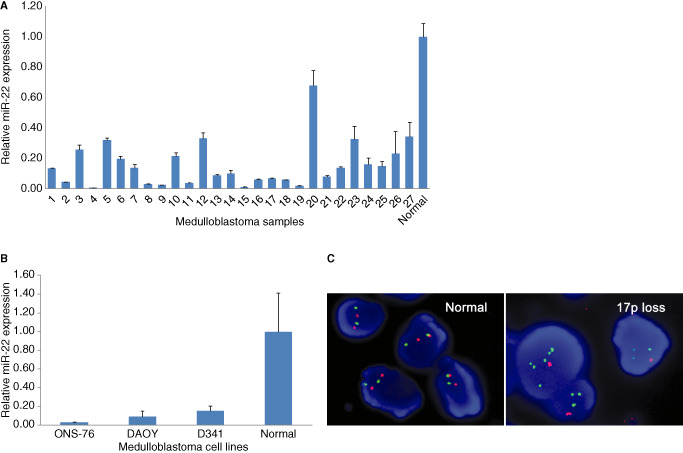
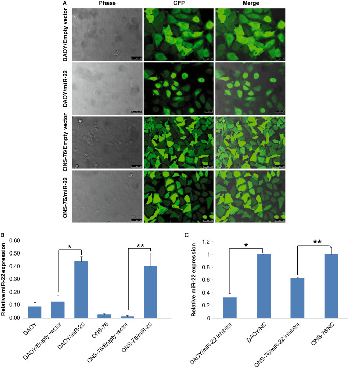
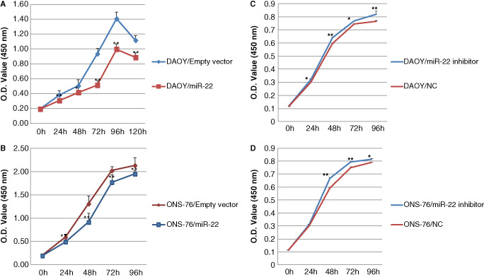
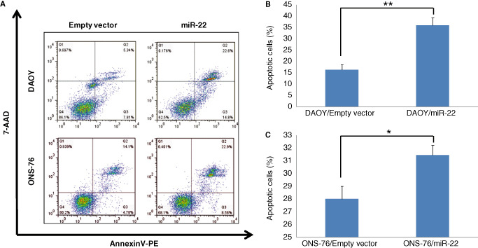
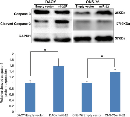
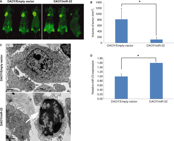
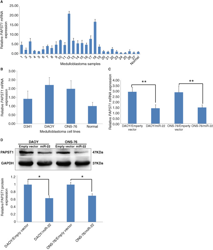
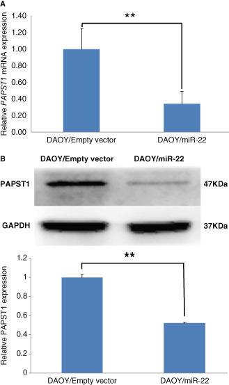
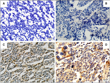
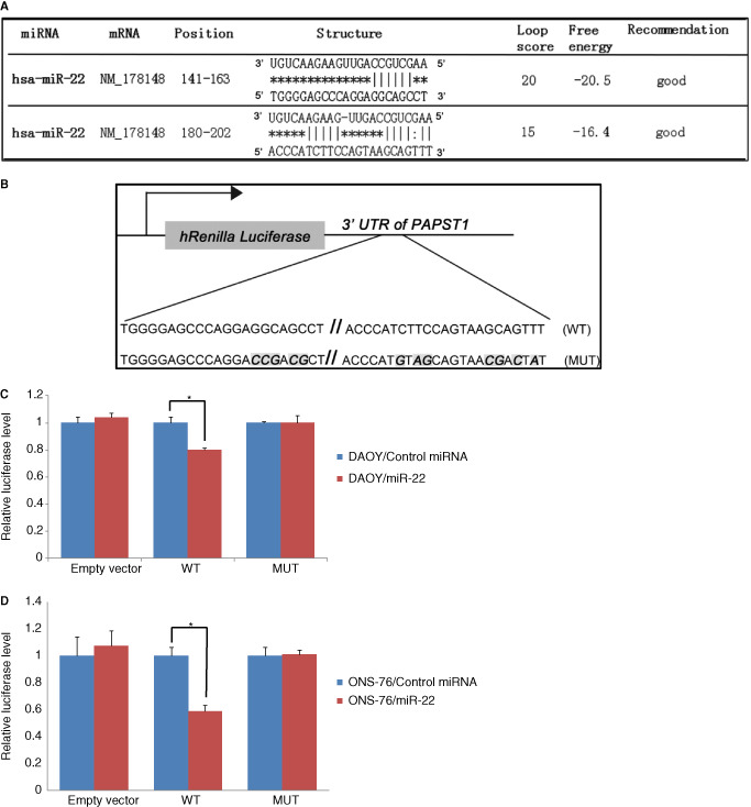
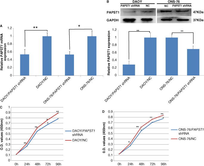
Medulloblastoma is the most frequent malignant central nervous system tumor in children. MicroRNAs (miRs) are small, non-coding RNAs that target protein-coding and non-coding RNAs, and play roles in a variety of cellular processes through regulation of multiple targets. In the present study, we analyzed miR-22 expression and its effect in cell proliferation and apoptosis in medulloblastomas. Quantitative reverse transcription PCR (RT-PCR) revealed significantly lower expression of miR-22 in 19 out of 27 (70%) medulloblastomas, D341, DAOY, ONS-76 medulloblastoma cell lines, compared with normal cerebellum. Forced expression of miR-22 by lentiviral vector transfection reduced cell proliferation and induced apoptosis, while knockdown of miR-22 increased proliferative activity in DAOY and ONS-76 cells. DAOY cells with miR-22 overexpression in nude mice yielded tumors smaller than those originated from control DAOY cells. Microarray analysis in DAOY cells with forced miR-22 expression showed significant changes in expression profiles, PAPST1 being the most significantly (10 folds) downregulated gene. Quantitative RT-PCR revealed PAPST1 mRNA upregulation in 18 out of 27 (67%) medulloblastomas. In addition, a luciferase reporter assay in ONS-76 and DAOY cells suggested that miR-22 directly targets the PAPST1 gene, and lentivirus-mediated knockdown of PAPST1 suppressed proliferation of DAOY and ONS-76 medulloblastoma cells. These results suggest that frequently downregulated miR-22 expression is associated with cell proliferation in medulloblastomas, and this may be at least in part via PAPST1, which is a novel target of miR-22.
Keywords: PAPST1; apoptosis; cell proliferation; medulloblastoma; miR-22.
© 2014 International Society of Neuropathology.
Figures
Similar articles
-
miR-124 is frequently down-regulated in medulloblastoma and is a negative regulator of SLC16A1.Hum Pathol. 2009 Sep;40(9):1234-43. doi: 10.1016/j.humpath.2009.02.003. Epub 2009 May 7. Hum Pathol. 2009. PMID: 19427019
-
Triptolide inhibits the proliferation and migration of medulloblastoma Daoy cells by upregulation of microRNA-138.J Cell Biochem. 2018 Dec;119(12):9866-9877. doi: 10.1002/jcb.27307. Epub 2018 Aug 28. J Cell Biochem. 2018. Retraction in: J Cell Biochem. 2021 Nov;122 Suppl 1:S24. doi: 10.1002/jcb.29983. PMID: 30156009 Retracted.
-
[MicroRNA383 regulates expression of PRDX3 in human medulloblastomas].Zhonghua Bing Li Xue Za Zhi. 2012 Aug;41(8):547-52. doi: 10.3760/cma.j.issn.0529-5807.2012.08.009. Zhonghua Bing Li Xue Za Zhi. 2012. PMID: 23157748 Chinese.
-
Genetic alterations in microRNAs in medulloblastomas.Brain Pathol. 2012 Mar;22(2):230-9. doi: 10.1111/j.1750-3639.2011.00523.x. Epub 2011 Sep 15. Brain Pathol. 2012. PMID: 21793975 Free PMC article.
-
MiR-206, a Cerebellum Enriched miRNA Is Downregulated in All Medulloblastoma Subgroups and Its Overexpression Is Necessary for Growth Inhibition of Medulloblastoma Cells.J Mol Neurosci. 2015 Jul;56(3):673-80. doi: 10.1007/s12031-015-0548-z. Epub 2015 Apr 10. J Mol Neurosci. 2015. PMID: 25859932
Cited by
-
Sweet Control: MicroRNA Regulation of the Glycome.Biochemistry. 2020 Sep 1;59(34):3098-3110. doi: 10.1021/acs.biochem.9b00784. Epub 2019 Oct 21. Biochemistry. 2020. PMID: 31585501 Free PMC article. Review.
-
MiR-22, regulated by MeCP2, suppresses gastric cancer cell proliferation by inducing a deficiency in endogenous S-adenosylmethionine.Oncogenesis. 2020 Nov 10;9(11):99. doi: 10.1038/s41389-020-00281-z. Oncogenesis. 2020. PMID: 33168819 Free PMC article.
-
MicroRNA-22: a Novel and Potent Biological Therapeutics in Neurological Disorders.Mol Neurobiol. 2022 May;59(5):2694-2701. doi: 10.1007/s12035-022-02769-8. Epub 2022 Feb 14. Mol Neurobiol. 2022. PMID: 35156160 Review.
-
Methods of miRNA delivery and possibilities of their application in neuro-oncology.Noncoding RNA Res. 2023 Oct 7;8(4):661-674. doi: 10.1016/j.ncrna.2023.10.002. eCollection 2023 Dec. Noncoding RNA Res. 2023. PMID: 37860265 Free PMC article. Review.
-
ShRNA-based POLD2 expression knockdown sensitizes glioblastoma to DNA-Damaging therapeutics.Cancer Lett. 2020 Jul 10;482:126-135. doi: 10.1016/j.canlet.2020.01.011. Epub 2020 Jan 16. Cancer Lett. 2020. PMID: 31954770 Free PMC article.
References
-
- Bai AH, Milde T, Remke M, Rolli CG, Hielscher T, Cho YJ et al (2012) MicroRNA‐182 promotes leptomeningeal spread of non‐sonic hedgehog‐medulloblastoma. Acta Neuropathol 123:529–538. - PubMed
Publication types
MeSH terms
Substances
Grants and funding
LinkOut - more resources
Full Text Sources
Other Literature Sources
Molecular Biology Databases
