

Host cell virus entry mediated by Australian bat lyssavirus G envelope glycoprotein occurs through a clathrin-mediated endocytic pathway that requires actin and Rab5
- PMID: 24576301
- PMCID: PMC3946599
- DOI: 10.1186/1743-422X-11-40
Host cell virus entry mediated by Australian bat lyssavirus G envelope glycoprotein occurs through a clathrin-mediated endocytic pathway that requires actin and Rab5
Abstract
Background: Australian bat lyssavirus (ABLV), a rhabdovirus of the genus Lyssavirus which circulates in both pteropid fruit bats and insectivorous bats in mainland Australia, has caused three fatal human infections, the most recent in February 2013, manifested as acute neurological disease indistinguishable from clinical rabies. Rhabdoviruses infect host cells through receptor-mediated endocytosis and subsequent pH-dependent fusion mediated by their single envelope glycoprotein (G), but the specific host factors and pathways involved in ABLV entry have not been determined.
Methods: ABLV internalization into HEK293T cells was examined using maxGFP-encoding recombinant vesicular stomatitis viruses (rVSV) that express ABLV G glycoproteins. A combination of chemical and molecular approaches was used to investigate the contribution of different endocytic pathways to ABLV entry. Dominant negative Rab GTPases were used to identify the endosomal compartment utilized by ABLV to gain entry into the host cell cytosol.
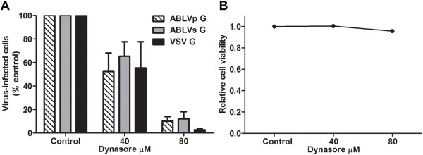
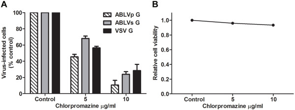
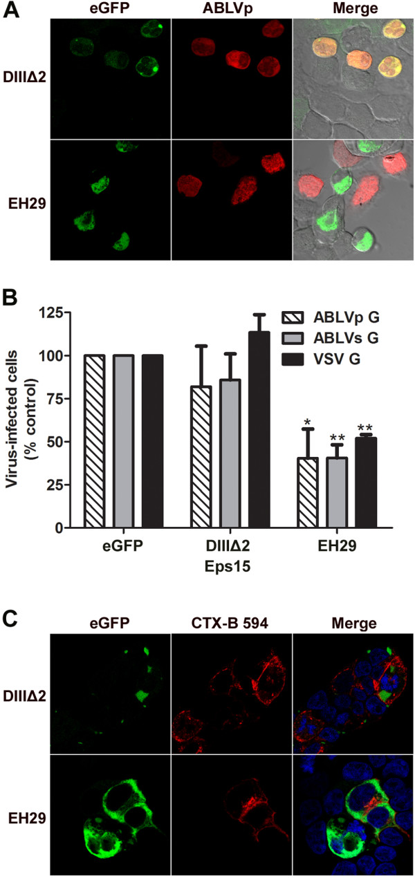
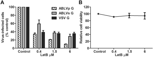
Results: Here we show that ABLV G-mediated entry into HEK293T cells was significantly inhibited by the dynamin-specific inhibitor dynasore, chlorpromazine, a drug that blocks clathrin-mediated endocytosis, and the actin depolymerizing drug latrunculin B. Over expression of dominant negative mutants of Eps15 and Rab5 also significantly reduced ABLV G-mediated entry into HEK293T cells. Chemical inhibitors of caveolae-dependent endocytosis and macropinocytosis and dominant negative mutants of Rab7 and Rab11 had no effect on ABLV entry.
Conclusions: The predominant pathway utilized by ABLV for internalization into HEK293T cells is clathrin-and actin-dependent. The requirement of Rab5 for productive infection indicates that ABLV G-mediated fusion occurs within the early endosome compartment.
Figures
Similar articles
-
Host cell tropism mediated by Australian bat lyssavirus envelope glycoproteins.Virology. 2013 Sep;444(1-2):21-30. doi: 10.1016/j.virol.2013.06.016. Epub 2013 Jul 10. Virology. 2013. PMID: 23849788 Free PMC article.
-
Recent observations on Australian bat lyssavirus tropism and viral entry.Viruses. 2014 Feb 19;6(2):909-26. doi: 10.3390/v6020909. Viruses. 2014. PMID: 24556791 Free PMC article. Review.
-
Entry of Classical Swine Fever Virus into PK-15 Cells via a pH-, Dynamin-, and Cholesterol-Dependent, Clathrin-Mediated Endocytic Pathway That Requires Rab5 and Rab7.J Virol. 2016 Sep 29;90(20):9194-208. doi: 10.1128/JVI.00688-16. Print 2016 Oct 15. J Virol. 2016. PMID: 27489278 Free PMC article.
-
Rab5 and Rab11 Are Required for Clathrin-Dependent Endocytosis of Japanese Encephalitis Virus in BHK-21 Cells.J Virol. 2017 Sep 12;91(19):e01113-17. doi: 10.1128/JVI.01113-17. Print 2017 Oct 1. J Virol. 2017. PMID: 28724764 Free PMC article.
-
Emergence, Tropism, Disease, and Treatment of Australian Bat Lyssavirus Infections in Humans.Vector Borne Zoonotic Dis. 2023 Sep;23(9):486-494. doi: 10.1089/vbz.2022.0089. Epub 2023 Jun 19. Vector Borne Zoonotic Dis. 2023. PMID: 37335942 Review.
Cited by
-
Human, Nonhuman Primate, and Bat Cells Are Broadly Susceptible to Tibrovirus Particle Cell Entry.Front Microbiol. 2019 Apr 26;10:856. doi: 10.3389/fmicb.2019.00856. eCollection 2019. Front Microbiol. 2019. PMID: 31105663 Free PMC article.
-
Entry of Challenge Virus Standard (CVS) -11 into N2a cells via a clathrin-mediated, cholesterol-, dynamin-, pH-dependent endocytic pathway.Virol J. 2019 Jun 13;16(1):80. doi: 10.1186/s12985-019-1186-9. Virol J. 2019. PMID: 31196105 Free PMC article.
-
Rhabdovirus Infection Is Dependent on Serine/Threonine Kinase AP2-Associated Kinase 1.Life (Basel). 2020 Aug 30;10(9):170. doi: 10.3390/life10090170. Life (Basel). 2020. PMID: 32872567 Free PMC article.
-
Status of antiviral therapeutics against rabies virus and related emerging lyssaviruses.Curr Opin Virol. 2019 Apr;35:1-13. doi: 10.1016/j.coviro.2018.12.009. Epub 2019 Feb 10. Curr Opin Virol. 2019. PMID: 30753961 Free PMC article. Review.
-
ICAM-1 Binding Rhinoviruses A89 and B14 Uncoat in Different Endosomal Compartments.J Virol. 2016 Aug 12;90(17):7934-42. doi: 10.1128/JVI.00712-16. Print 2016 Sep 1. J Virol. 2016. PMID: 27334586 Free PMC article.
References
-
- Allworth A, Murray K, Morgan J. A human case of encephalitis due to a lyssavirus recently identified in fruit bats. Commun Dis Intellig. 1996;20:504.
-
- Australian bat lyssavirus - Australia (02): Queensland, human fatality. ProMed-mail (International Society for Infectious Diseases, 21 March 2013, archive no. 20130323.1600266); http://www.promedmail.org.
-
- Hanna JN, Carney IK, Smith GA, Tannenberg AE, Deverill JE, Botha JA, Serafin IL, Harrower BJ, Fitzpatrick PF, Searle JW. Australian bat lyssavirus infection: a second human case, with a long incubation period. Med J Aust. 2000;172:597–599. - PubMed
-
- Australian bat lyssavirus - Australia (04): Equine Fatalities. ProMed-mail (International Society for Infectious Diseases, 17 May 2013, archive no. 20130517.1720540); http://www.promedmail.org.
Publication types
MeSH terms
Substances
Grants and funding
LinkOut - more resources
Full Text Sources
Other Literature Sources
Research Materials
Miscellaneous
