

Proteomic identification of fucosylated haptoglobin alpha isoforms in ascitic fluids and its localization in ovarian carcinoma tissues from Mexican patients
- PMID: 24576319
- PMCID: PMC3943579
- DOI: 10.1186/1757-2215-7-27
Proteomic identification of fucosylated haptoglobin alpha isoforms in ascitic fluids and its localization in ovarian carcinoma tissues from Mexican patients
Abstract
Background: Ovarian cancer is the most lethal gynecologic disease due to delayed diagnosis, and ascites production is a characteristic of patients in advanced stages. The aim of this study was to perform the proteomic analysis of ascitic fluids of Mexican patients with ovarian carcinoma, in order to detect proteins with a differential expression pattern in the continuing search to identify biomarkers for this disease.
Methods: Samples were collected from 50 patients from the Instituto Nacional de Cancerología of México under informed consent and with approval of the bioethics and scientific committees. After elimination of abundant proteins (Albumin/IgGs) samples were processed for 2D electrophoresis and further protein identification by Mass Spectrometry (MALDI-TOF). Molecules of interest were followed by western blot and lectin binding assays, and their tissue location by histo-immunofluorescence and confocal analysis.
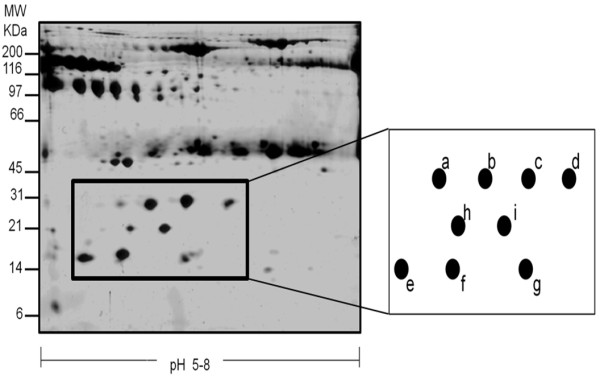
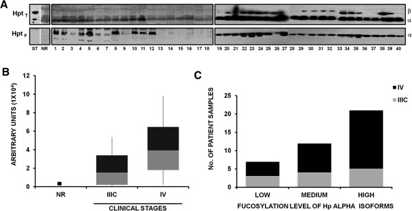
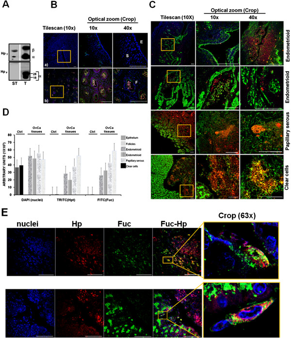
Results and discussion: An area with a differential expression pattern among samples was located in the 2D gels. Identified proteins were 6 alpha 1 isoforms and 1 alpha 2 isoform of Haptoglobin, and 2 isoforms of Transthyretin. While Transthyretin isoforms were constitutively expressed in all samples, clear differences in the expression pattern of Haptoglobin alpha isoforms were found. Moreover, increased levels of fucosylation of Haptoglobin alpha isoforms analyzed in 40 samples by Aleuria aurantia lectin binding by 1D overlay assay showed a positive correlation with advanced stages of the disease. Tissue detection of Haptoglobin and its fucosylated form, by histo-immunofluorescence in biopsies of ovarian cancer, also showed a correlation with ovarian cancer progression. Moreover, results show that fucosylated Haptoglobin is produced by tumor cells.
Conclusions: Increased numbers of highly fucosylated Haptoglobin alpha isoforms in ascitic fluids and the presence of fucosylated Haptoglobin in tumor tissues of ovarian cancer Mexican patients associated with advanced stages of the disease, reinforce the potential of fucosylated Haptoglobin alpha isoforms to be characterized as biomarkers for disease progression.
Figures

Similar articles
-
Integrins and haptoglobin: Molecules overexpressed in ovarian cancer.Pathol Res Pract. 2015 Dec;211(12):973-81. doi: 10.1016/j.prp.2015.10.002. Epub 2015 Nov 6. Pathol Res Pract. 2015. PMID: 26549463
-
Proteomic-based identification of haptoglobin-1 precursor as a novel circulating biomarker of ovarian cancer.Br J Cancer. 2004 Jul 5;91(1):129-40. doi: 10.1038/sj.bjc.6601882. Br J Cancer. 2004. PMID: 15199385 Free PMC article.
-
Identification of oncofetal fibronectin in patients with advanced epithelial ovarian cancer: detection in ascitic fluid and localization to primary sites and metastatic implants.Cancer. 1998 Jan 1;82(1):152-8. doi: 10.1002/(sici)1097-0142(19980101)82:1<152::aid-cncr19>3.0.co;2-1. Cancer. 1998. PMID: 9428492
-
Fucosylated haptoglobin is a novel marker for pancreatic cancer: detailed analyses of oligosaccharide structures.Proteomics. 2008 Aug;8(16):3257-62. doi: 10.1002/pmic.200800046. Proteomics. 2008. PMID: 18646007 Review.
-
Identification of fucosylated haptoglobin as a novel tumor marker for pancreatic cancer and its possible application for a clinical diagnostic test.Methods Enzymol. 2010;478:153-64. doi: 10.1016/S0076-6879(10)78006-X. Methods Enzymol. 2010. PMID: 20816478 Review.
Cited by
-
Haptoglobin and CCR2 receptor expression in ovarian cancer cells that were exposed to ascitic fluid: exploring a new role of haptoglobin in the tumoral microenvironment.Cell Adh Migr. 2015;9(5):394-405. doi: 10.1080/19336918.2015.1035504. Epub 2015 Jul 25. Cell Adh Migr. 2015. PMID: 26211665 Free PMC article.
-
Improvement of core-fucosylated glycoproteome coverage via alternating HCD and ETD fragmentation.J Proteomics. 2016 Sep 2;146:90-8. doi: 10.1016/j.jprot.2016.06.003. Epub 2016 Jun 6. J Proteomics. 2016. PMID: 27282921 Free PMC article.
-
Haptoglobin as a Biomarker.Biochem Mosc Suppl B Biomed Chem. 2021;15(3):184-198. doi: 10.1134/S1990750821030069. Epub 2021 Aug 16. Biochem Mosc Suppl B Biomed Chem. 2021. PMID: 34422226 Free PMC article.
-
Construction of 2DE Patterns of Plasma Proteins: Aspect of Potential Tumor Markers.Int J Mol Sci. 2022 Sep 21;23(19):11113. doi: 10.3390/ijms231911113. Int J Mol Sci. 2022. PMID: 36232415 Free PMC article.
-
Evaluation of Haptoglobin and Its Proteoforms as Glioblastoma Markers.Int J Mol Sci. 2021 Jun 18;22(12):6533. doi: 10.3390/ijms22126533. Int J Mol Sci. 2021. PMID: 34207114 Free PMC article.
References
-
- Lamendola DE, Duan Z, Yusuf RZ, Seiden MV. Molecular description of evolving paclitaxel resistance in the SKOV-3 human ovarian carcinoma cell line. Cancer Res. 2003;63:2200–2205. - PubMed
Publication types
MeSH terms
Substances
LinkOut - more resources
Full Text Sources
Other Literature Sources
Medical
Research Materials
Miscellaneous
