. 2014 Jun;1843(6):1196-206.
doi: 10.1016/j.bbamcr.2014.02.008. Epub 2014 Feb 24.

Specificity protein 4 (Sp4) regulates the transcription of AMPA receptor subunit GluA2 (Gria2)

Affiliations

Specificity protein 4 (Sp4) regulates the transcription of AMPA receptor subunit GluA2 (Gria2)

Anusha Priya et al. Biochim Biophys Acta. 2014 Jun.

Abstract

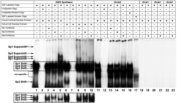
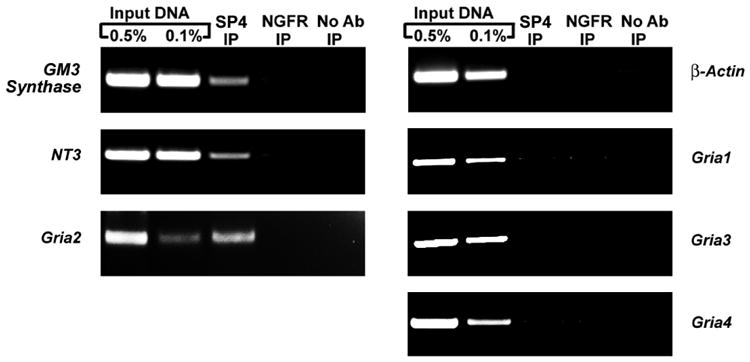
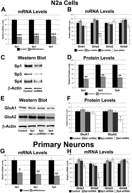
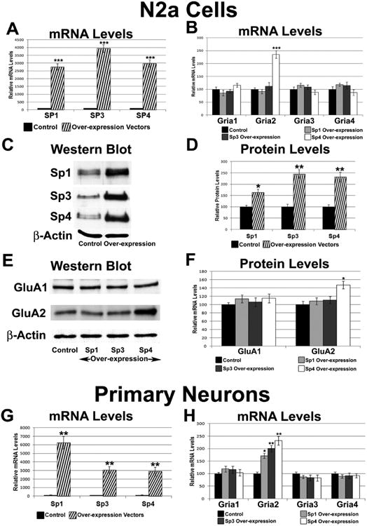
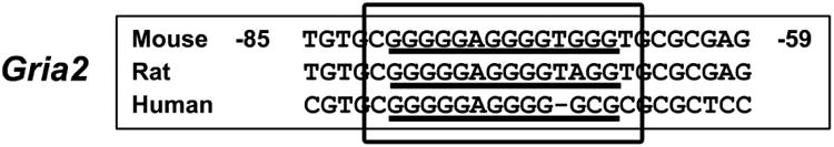
The alpha-amino-3-hydroxy-5-methyl-4-isoxazolepropionic acid (AMPA) receptors are important glutamatergic receptors mediating fast excitatory synaptic transmission in the brain. The regulation of the four subunits of AMPA receptors, GluA1-4, is poorly understood. Excitatory synaptic transmission is highly energy-demanding, and this energy is derived mainly from the oxidative pathway. Recently, we found that specificity factor regulates all subunits of cytochrome c oxidase (COX), a critical energy-generating enzyme. COX is also regulated by nuclear respiratory factor 1 (NRF-1), which transcriptionally controls the Gria2 (GluA2) gene of AMPA receptors. The goal of the present study was to test our hypothesis that Sp-factors (Sp1, Sp3, and/or Sp4) also regulate AMPA subunit genes. If so, we wish to determine if Sp-factors and NRF-1 function via a complementary, concurrent and parallel, or a combination of complementary and concurrent/parallel mechanism. By means of multiple approaches, including electrophoretic mobility shift and supershift assays, chromatin immunoprecipitation, promoter mutations, real-time quantitative PCR, and western blot analysis, we found that Sp4, but not Sp1 or Sp3, regulates the Gria2, but not Gria1, 3, or 4, subunit gene of the AMPA receptor in a concurrent and parallel manner with NRF-1. Thus, Sp4 and NRF-1 both mediate the tight coupling between neuronal activity and energy metabolism at the transcriptional level.

Keywords: AMPA receptor; Gene regulation; GluA2; Sp4; Specificity protein 4; Transcription factor.


Conflict of interest statement

All authors have no conflict of interest to declare.

Figures

Figure 1
Figure 1
In vitro binding of Sp factors to the GM3 Synthase and Gria2 promoters using EMSA and supershift assays. All lanes contain 32P-labeled oligonucleotides and are labeled by a “+” or a “−“sign depending on whether they also contain mouse visual cortical or HeLa nuclear extract, excess oligos or mutant oligos (unlabeled), and Sp1, Sp3, or Sp4 antibodies. Sp1, Sp3, or Sp4 shift, supershift, and non-specific complexes are indicated by arrows. The positive control for Sp factor binding was GM3 Synthase. Specific Sp1, 3, or 4 shift bands were revealed upon incubation with cortical (lanes 1-5) or HeLa (lanes 5-10) nuclear extract (lanes 1 and 6, respectively). Excess unlabeled competitor competed the shift bands (lanes 2 and 7). The addition of Sp1 antibody did not yield a supershift band with cortical nuclear extract (lane 3) but gave two specific supershift bands with HeLa nuclear extract (lane 8), corresponding to the presence of tandem Sp binding. The addition of Sp3 antibody yielded a faint specific supershift band with cortical nuclear extract (lane 4) and a stronger supershift band with HeLa nuclear extract (lane 9). The addition of Sp4 antibody yielded a very strong supershift band with cortical nuclear extract (lane 5) and a much fainter supershift band with HeLa nuclear extract (lane 10). The relative levels of the Sp1, Sp3, and Sp4 supershift bands in mouse cortical and HeLa nuclear extract corresponds to the relative levels of these transcription factors in these tissue and cell types. As Sp1, 3, and 4 all recognize the same cis motif, their combined binding yielded a rather thick shift band. However, with shorter gel exposure (boxed lanes 1 to 10 highlighted in the lower left), the individual shift bands can sometimes be dissociated. As shown in lane 1 in the inset, cortical nuclear extract did not reveal any detectable Sp1 shift band, but did show distinct Sp3 and Sp4 bands. The Sp3 shift band disappeared when it was supershifted (lane 4). Likewise, the Sp4 shift band disappeared when it was supershifted (lane 5). With HeLa nuclear extract, the rather abundant presence of both Sp1 and Sp3 caused the bands to merge even with lighter exposure (lane 6). However, the upper portion (Sp1) disappeared when supershifted with Sp1 antibody (lane 8), and the lower portion (Sp3) became lighter when supershifted with Sp3 antibody (lane 9). No change was detectable with anti-Sp4 antibody (lane 10), indicating that HeLa nuclear extract did not contain detectable Sp4. Incubation of cortical nuclear extract with the Gria2 probe yielded a specific Sp4 shift band (lane 11) which was competed out with the addition of excess cold probes (lane 12). The addition of Sp4 anitbody yielded a specific supershift band (lane 15), but the addition of Sp1 and Sp3 antibodies did not (lanes 13 and 14). The addition of excess unlabeled mutant Gria2 probes did not compete out the shift reaction (lane 16). Labeled Gria2 probes with mutant Sp sites did not yield specific Sp shift bands (lane 17). Labeled Gria1, Gria3, and Gria4 probes did not yield specific Sp shift (lanes 18, 20, and 22, respectively) or Sp4 supershift bands (lanes 19, 21, and 23, respectively).
Figure 2
Figure 2
Sp4 interactions with AMPA receptor subunit promoters in mouse visual cortical tissue. Precipitation of chromatin was carried out from mouse visual cortical tissue nuclear extract with anti Sp4 antibodies (Sp4 IP lane), anti-nerve growth factor receptor p75 antibody (negative control, NGFR IP lane), or no antibody (negative control, No Ab lane). 0.5% (input 0.5% IP lane) and 0.1% (input 0.1% IP lane) of input chromatin served as control reactions for PCR. Positive controls for Sp4 binding were GM3 synthase and Neurotrophin 3, whereas p-Actin was the negative control. Sp4 interacted with Gria2, but not with Gria1, Gria3, or Gria4.
Figure 3
Figure 3
Relative luciferase activity of the wild type Gria2 promoter (wt) and the Gria2 promoter with mutated Sp binding site (mut). Mutating the Sp binding site on Gria2 resulted in a significant decrease in luciferase activity as compared to the wild type promoter. KCl depolarization significantly increased Gria2 wild type promoter activity, but could not increase activity in the Gria2 promoter with mutated Sp site. N = 6 for each construct. ***= P < 0.001; X = NS. Both mutant and wild type + KCl were compared to the wild type. The mutant + KCl was compared to the mutant.
Figure 4
Figure 4
Effect of silencing of Sp1, Sp3, or Sp4 on the expression of the AMPA receptor subunit genes using RNA interference. (A) N2a cells transfected with Sp1, Sp3, or Sp4 shRNA showed significant down-regulation of Sp1, Sp3, and Sp4 transcripts, respectively. N = 6. (B) In N2a cells, Gria2 transcript levels were decreased with silencing of Sp4, but not with silencing of either Sp3 or Sp1. N = 6. (C-D) In N2a cells, protein levels of Sp1, Sp3, and Sp4 decreased significantly with shRNA against Sp1, Sp3, and Sp4, respectively. A representative western blot for β-actin, the loading control, is shown. N = 3. (E-F) Sp4 silencing reduced the protein levels of GluA2 but not that of GluA1. Protein levels of GluA1 and GluA2 did not change significantly with Sp1 or Sp3 silencing. The loading control was β-actin. N = 3. (G) shRNA against Sp1, Sp3, or Sp4 showed a significant down-regulation in the transcript levels of Sp1, Sp3, and Sp4, respectively. N = 3. (H) Transcript levels of Gria2, but not Gria1, Gria3, and Gria4, decreased with Sp4 shRNA in primary neurons. Transcript levels of Gria1-4 did not change significantly with Sp1 or Sp3 shRNA in primary neurons. N = 3. *= P < 0.05, **= P < 0.01, and ***= P < 0.001 when compared to controls.
Figure 5
Figure 5
Effect of over-expressing Sp1, Sp3, or Sp4 on the transcript levels of AMPA receptor subunit genes. (A) N2a cells transfected with Sp1, Sp3, and Sp4 expression vectors revealed an up-regulation of Sp1, Sp3, and Sp4 transcripts, respectively. N = 6. (B) Sp4 over-expression, but not Sp1 or Sp3 over-expression, increased Gria2 transcript levels. N = 6. (C-D) In N2a cells, protein levels of Sp1, Sp3, and Sp4 were increased significantly with over-expression of Sp1, Sp3, and Sp4, respectively. A representative blot of the loading control, β-actin, is shown. N = 3. (E-F) In N2a cells, the protein level of GluA2, but not that of GluA1, was increased significantly with over-expression of Sp4. Protein levels of GluA1 and GluA2 did not change significantly with over-expressing Sp1 or Sp3. The loading control was β-actin. N = 3. (G) Over-expression of Sp1, Sp3, or Sp4 in primary neurons increased transcript levels of Sp1, Sp3, and Sp4, respectively. N = 3. (H) Sp4, Sp3, and Sp1 over-expression increased Gria2 transcript levels, but not Gria1, Gria3 and Gria4 transcript levels, in primary neurons. N = 3. *= P < 0.05, **= P < 0.01, and ***= P < 0.001 when compared to controls.
Figure 6
Figure 6
Effect of changes in neuronal activity on Gria1, 2, 3, and 4 transcript levels in the absence or presence of Sp1, Sp3, or Sp4 silencing or over-expression in N2a cells. (A) Transcript levels of all AMPA receptor subunit genes were increased by 5 h of 20 mM KCl treatment. KCl is a depolarizing agent that increases neuronal activity. Silencing Sp4 did not allow for transcript levels of Gria2 to increase with KCl, but had no effect on the KCl-induced increase in the transcript levels of Gria1, Gria3, and Gria4. Silencing Sp1 and Sp3 did not prevent the KCl-induced up-regulation of Gria1-4 transcript levels. N = 6. (B) Transcript levels of all AMPA receptor subunit genes were decreased with 3 days of 0.4 μM TTX treatment. TTX decreases neuronal activity by blocking action potentials. Sp4 over-expression rescued the TTX-mediated down-regulation of Gria2 transcript levels, but did not rescue the TTX-mediated down-regulation of the Gria1, Gria3, and Gria4 transcripts. Sp1 or Sp3 over-expression could not rescue the TTX-mediated down-regulation of Gria1-4 transcript levels. N = 6. *= P < 0.05, **= P < 0.01, and ***= P < 0.001 when compared to controls. ### = P < 0.001 and X = non-significant when compared to KCl or TTX treatment alone.
Figure 7
Figure 7
Effect of changes in neuronal activity on Gria1, 2, 3, and 4 transcript levels in the absence or presence of Sp4 silencing or over-expression in rat visual cortical neurons. (A) Transcript levels of Sp4 and all AMPA receptor subunit genes were increased by 5 h of 20 mM KCl treatment. Silencing Sp4 reduced its own transcript levels as well as that of Gria2, but not those of Gria1, 3, or 4. Sp4 silencing also did not allow for transcript levels of Sp4 itself and Gria2 to increase with KCl, but it had no effect on KCl-induced up-regulation of Gria1, Gria3, and Gria4 transcripts. N = 3. (B) Transcript levels of Sp4 and all AMPA receptor subunit genes were decreased with 3 days of 0.4 μM TTX treatment. Sp4 over-expression significantly increased its own transcript levels as well as that of Gria2, but not those of Gria1, 3, or 4. Sp4 over-expression also rescued the TTX-mediated down-regulation of Gria2 transcript levels, but not those of Gria1, Gria3, and Gria4 transcripts. N = 3. *= P < 0.05, **= P < 0.01, and ***= P < 0.001 when compared to controls. # = P < 0.05, ## = P < 0.01, ### = P < 0.001 and X = non-significant when compared to KCl or TTX treatment alone.
Figure 8
Figure 8
Aligned partial sequences of the Gria2 promoters from mice, rats, and humans showed conserved Sp binding sites.

Similar articles

Cited by

References

    1. Isaac JT, Ashby MC, McBain CJ. The role of the GluR2 subunit in AMPA receptor function and synaptic plasticity. Neuron. 2007;54:859–871. - PubMed
    1. Borges K, Dingledine R. AMPA receptors: molecular and functional diversity. Prog Brain Res. 1998;116:153–170. - PubMed
    1. Malinow R, Malenka RC. AMPA receptor trafficking and synaptic plasticity. Annu Rev Neurosci. 2002;25:103–126. - PubMed
    1. Turrigiano GG. The self-tuning neuron: synaptic scaling of excitatory synapses. Cell. 2008;135:422–435. - PMC - PubMed
    1. Shepherd JD, Huganir RL. The cell biology of synaptic plasticity: AMPA receptor trafficking. Annu Rev Cell Dev Biol. 2007;23:613–643. - PubMed

Publication types

MeSH terms