

TCDD induces dermal accumulation of keratinocyte-derived matrix metalloproteinase-10 in an organotypic model of human skin
- PMID: 24576722
- PMCID: PMC4137792
- DOI: 10.1016/j.taap.2014.02.010
TCDD induces dermal accumulation of keratinocyte-derived matrix metalloproteinase-10 in an organotypic model of human skin
Abstract
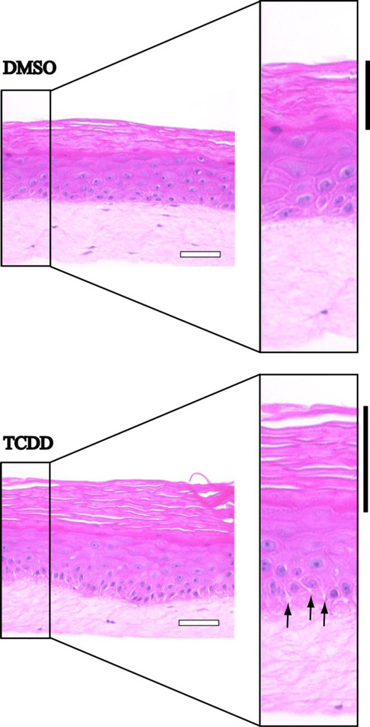
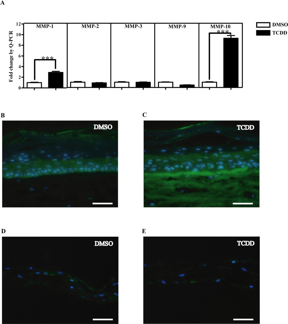
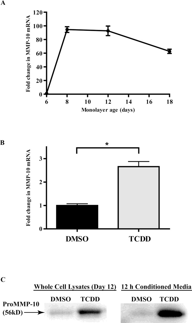
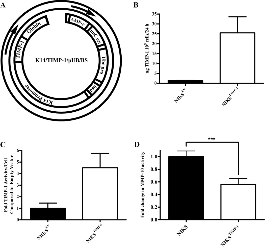
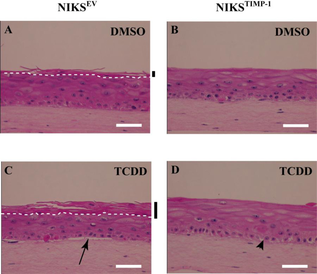
The epidermis of skin is the first line of defense against the environment. A three dimensional model of human skin was used to investigate tissue-specific phenotypes induced by the environmental contaminant, 2,3,7,8-tetrachlorodibenzo-p-dioxin (TCDD). Continuous treatment of organotypic cultures of human keratinocytes with TCDD resulted in intracellular spaces between keratinocytes of the basal and immediately suprabasal layers as well as thinning of the basement membrane, in addition to the previously reported hyperkeratinization. These tissue remodeling events were preceded temporally by changes in expression of the extracellular matrix degrading enzyme, matrix metalloproteinase-10 (MMP-10). In organotypic cultures MMP-10 mRNA and protein were highly induced following TCDD treatment. Q-PCR and immunoblot results from TCDD-treated monolayer cultures, as well as indirect immunofluorescence and immunoblot analysis of TCDD-treated organotypic cultures, showed that MMP-10 was specifically contributed by the epidermal keratinocytes but not the dermal fibroblasts. Keratinocyte-derived MMP-10 protein accumulated over time in the dermal compartment of organotypic cultures. TCDD-induced epidermal phenotypes in organotypic cultures were attenuated by the keratinocyte-specific expression of tissue inhibitor of metalloproteinase-1, a known inhibitor of MMP-10. These studies suggest that MMP-10 and possibly other MMP-10-activated MMPs are responsible for the phenotypes exhibited in the basement membrane, the basal keratinocyte layer, and the cornified layer of TCDD-treated organotypic cultures. Our studies reveal a novel mechanism by which the epithelial-stromal microenvironment is altered in a tissue-specific manner thereby inducing structural and functional pathology in the interfollicular epidermis of human skin.
Keywords: Dermis; Keratinocytes; MMP-10; NIKS; TCDD; TIMP-1.
Copyright © 2014 Elsevier Inc. All rights reserved.
Conflict of interest statement
None to declare.
Figures
Similar articles
-
2,3,7,8-Tetrachlorodibenzo-p-dioxin alters the differentiation pattern of human keratinocytes in organotypic culture.Toxicol Appl Pharmacol. 2001 Sep 1;175(2):121-9. doi: 10.1006/taap.2001.9202. Toxicol Appl Pharmacol. 2001. PMID: 11543644
-
Interaction between the aryl hydrocarbon receptor and retinoic acid pathways increases matrix metalloproteinase-1 expression in keratinocytes.J Biol Chem. 2004 Jun 11;279(24):25284-93. doi: 10.1074/jbc.M402168200. Epub 2004 Apr 9. J Biol Chem. 2004. PMID: 15075337
-
Influence of 2,3,7,8-tetrachlorodibenzo-p-dioxin (TCDD) on TNF-alpha levels in the skin of congenic haired and hairless mice.Toxicol Appl Pharmacol. 1994 Nov;129(1):12-5. doi: 10.1006/taap.1994.1223. Toxicol Appl Pharmacol. 1994. PMID: 7974484
-
Three-Dimensional Organotypic Systems for Modelling and Understanding Molecular Regulation of Oral Dentogingival Tissues.Int J Mol Sci. 2024 Oct 28;25(21):11552. doi: 10.3390/ijms252111552. Int J Mol Sci. 2024. PMID: 39519105 Free PMC article. Review.
-
Organotypic cultures as aging associated disease models.Aging (Albany NY). 2022 Nov 22;14(22):9338-9383. doi: 10.18632/aging.204361. Epub 2022 Nov 22. Aging (Albany NY). 2022. PMID: 36435511 Free PMC article. Review.
Cited by
-
Mitochondrial activity and oxidative stress functions are influenced by the activation of AhR-induced CYP1A1 overexpression in cardiomyocytes.Mol Med Rep. 2017 Jul;16(1):174-180. doi: 10.3892/mmr.2017.6580. Epub 2017 May 12. Mol Med Rep. 2017. PMID: 28498411 Free PMC article.
References
-
- Panteleyev AA, Bickers DR. Exp Dermatol. 2006;15:705–730. - PubMed
-
- Hudson LG, Toscano WA, Jr., Greenlee WF. Toxicol Appl Pharmacol. 1986;82:481–492. - PubMed
-
- Milstone LM, LaVigne JF. J Invest Dermatol. 1984;82:532–534. - PubMed
-
- Osborne R, Greenlee WF. Toxicol Appl Pharmacol. 1985;77:434–443. - PubMed
-
- Greenlee WF, Dold KM, Osborne R. In Vitro Cell Dev Biol. 1985;21:509–512. - PubMed
Publication types
MeSH terms
Substances
Grants and funding
LinkOut - more resources
Full Text Sources
Other Literature Sources
Research Materials
Miscellaneous
