

Acute 7,12-dimethylbenz[a]anthracene exposure causes differential concentration-dependent follicle depletion and gene expression in neonatal rat ovaries
- PMID: 24576726
- PMCID: PMC4077181
- DOI: 10.1016/j.taap.2014.02.011
Acute 7,12-dimethylbenz[a]anthracene exposure causes differential concentration-dependent follicle depletion and gene expression in neonatal rat ovaries
Abstract
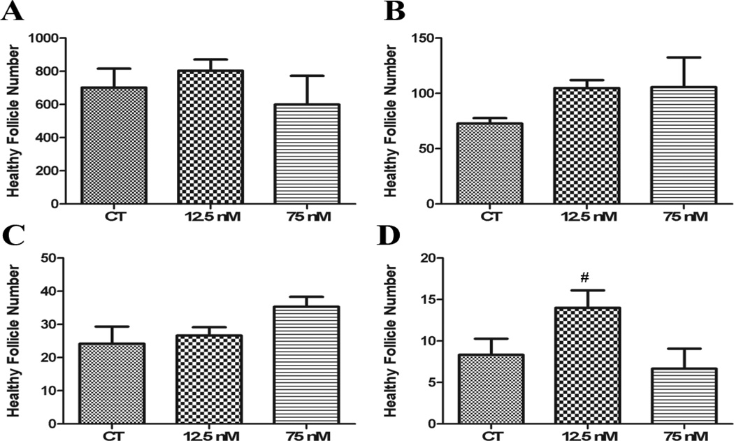
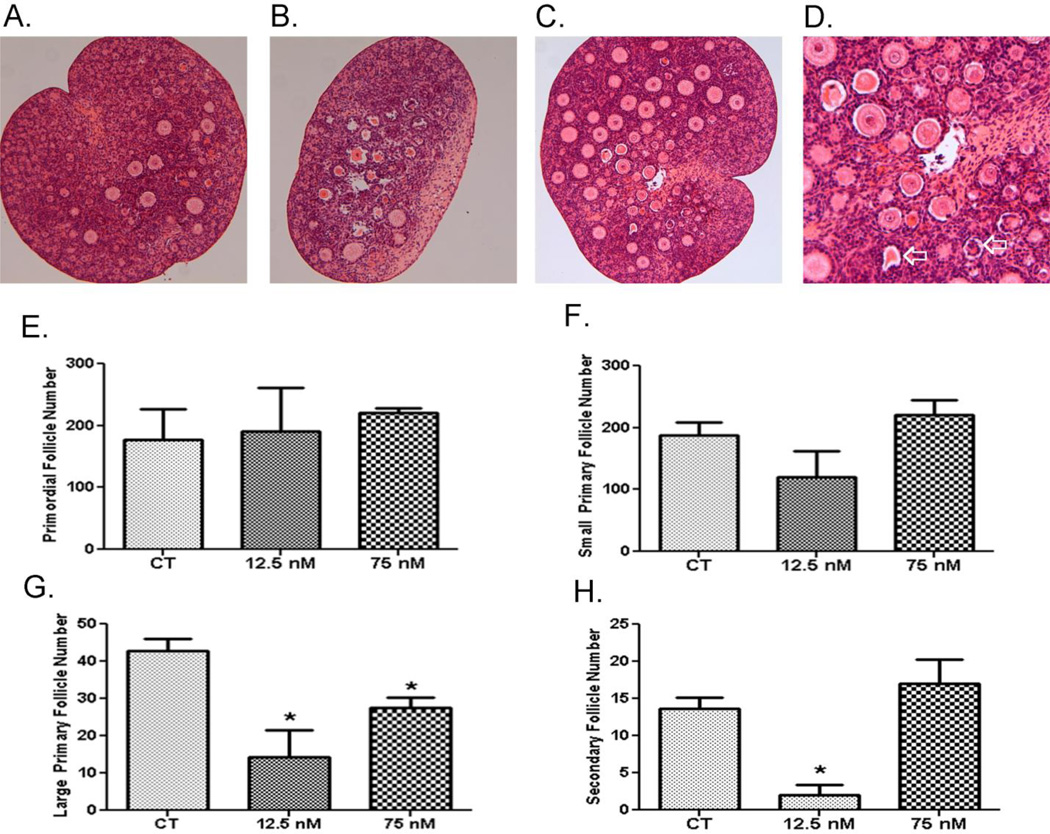
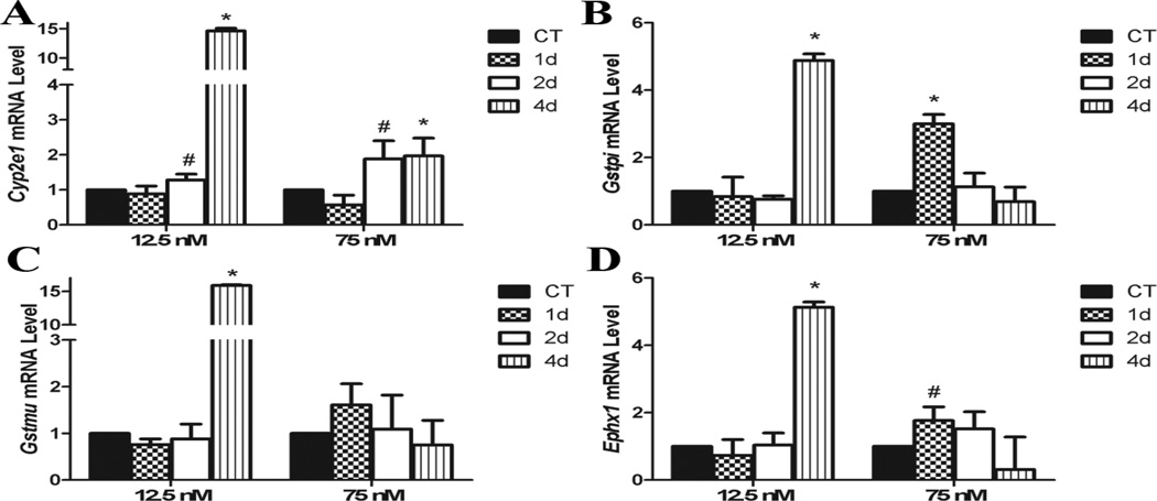
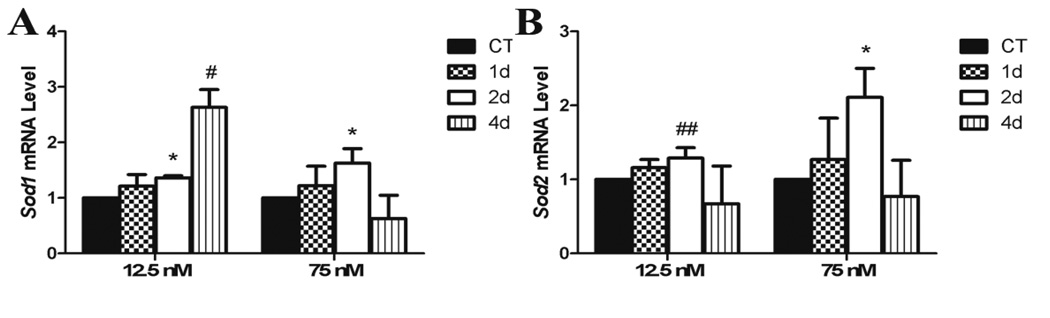
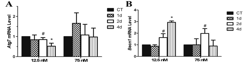
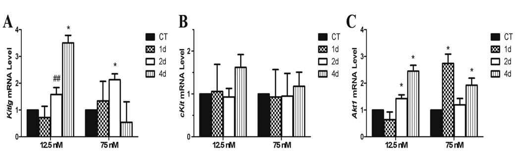
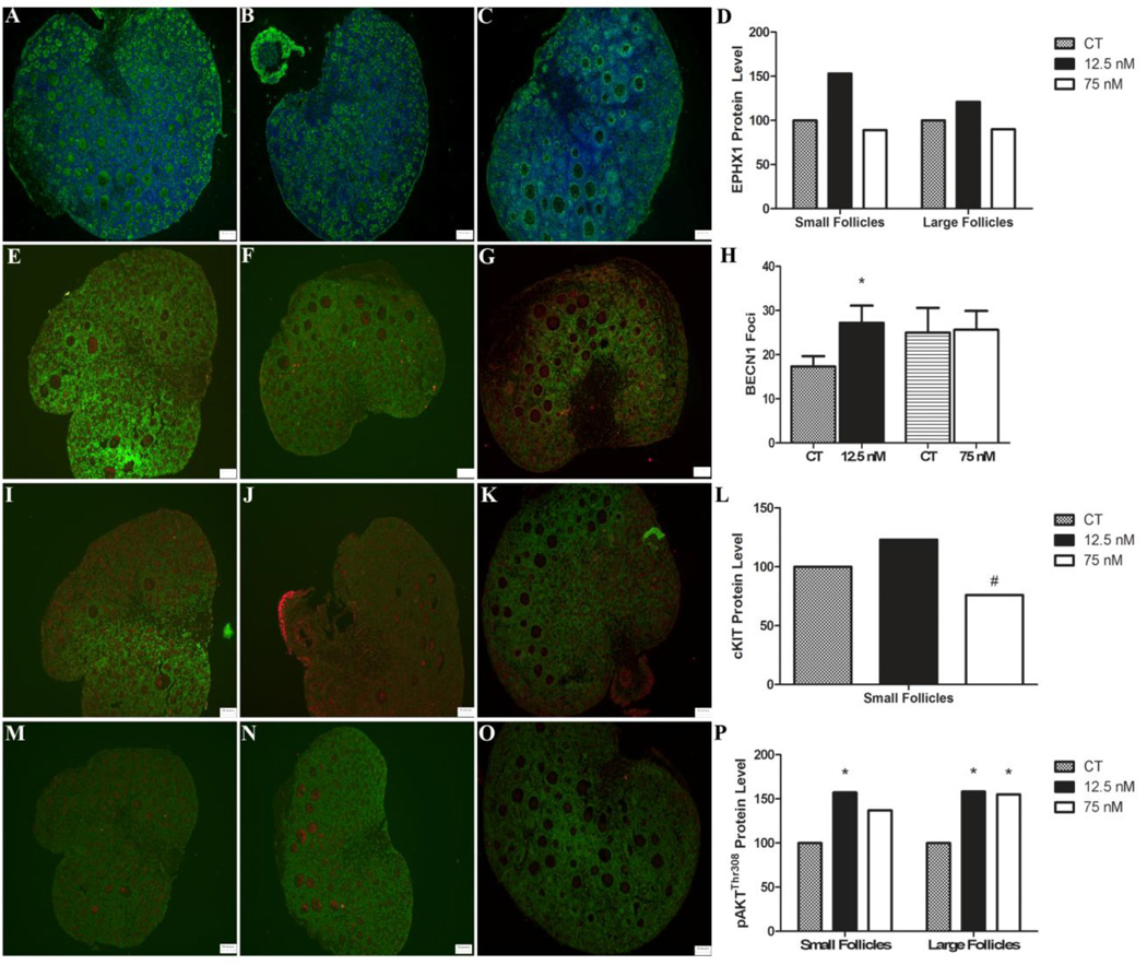
Chronic exposure to the polycyclic aromatic hydrocarbon 7,12-dimethylbenz[a]anthracene (DMBA), generated during combustion of organic matter including cigarette smoke, depletes all ovarian follicle types in the mouse and rat, and in vitro models mimic this effect. To investigate the mechanisms involved in follicular depletion during acute DMBA exposure, two concentrations of DMBA at which follicle depletion has (75 nM) and has not (12.5 nM) been observed were investigated. Postnatal day four F344 rat ovaries were maintained in culture for four days before a single exposure to vehicle control (1% DMSO; CT) or DMBA (12 nM; low-concentration or 75 nM; high-concentration). After four or eight additional days of culture, DMBA-induced follicle depletion was evaluated via follicle enumeration. Relative to control, DMBA did not affect follicle numbers after 4 days of exposure, but induced large primary follicle loss at both concentrations after 8 days; while, the low-concentration DMBA also caused secondary follicle depletion. Neither concentration affected primordial or small primary follicle number. RNA was isolated and quantitative RT-PCR performed prior to follicle loss to measure mRNA levels of genes involved in xenobiotic metabolism (Cyp2e1, Gstmu, Gstpi, Ephx1), autophagy (Atg7, Becn1), oxidative stress response (Sod1, Sod2) and the phosphatidylinositol 3-kinase (PI3K) pathway (Kitlg, cKit, Akt1) 1, 2 and 4 days after exposure. With the exception of Atg7 and cKit, DMBA increased (P < 0.05) expression of all genes investigated. Also, BECN1 and pAKT(Thr308) protein levels were increased while cKIT was decreased by DMBA exposure. Taken together, these results suggest an increase in DMBA bioactivation, add to the mechanistic understanding of DMBA-induced ovotoxicity and raise concern regarding female low concentration DMBA exposures.
Keywords: DMBA; Follicle; Ovary.
Copyright © 2014 Elsevier Inc. All rights reserved.
Figures
Similar articles
-
Evaluation of ovotoxicity induced by 7, 12-dimethylbenz[a]anthracene and its 3,4-diol metabolite utilizing a rat in vitro ovarian culture system.Toxicol Appl Pharmacol. 2009 Feb 1;234(3):361-9. doi: 10.1016/j.taap.2008.10.009. Epub 2008 Nov 5. Toxicol Appl Pharmacol. 2009. PMID: 19027032 Free PMC article.
-
Impact of obesity on ovotoxicity induced by 7,12-dimethylbenz[a]anthracene in mice.Biol Reprod. 2014 Mar 27;90(3):68. doi: 10.1095/biolreprod.113.114215. Print 2014 Mar. Biol Reprod. 2014. PMID: 24501177 Free PMC article.
-
7,12-Dimethylbenz[a]anthracene exposure induces the DNA repair response in neonatal rat ovaries.Toxicol Appl Pharmacol. 2013 Nov 1;272(3):690-6. doi: 10.1016/j.taap.2013.08.013. Epub 2013 Aug 19. Toxicol Appl Pharmacol. 2013. PMID: 23969067 Free PMC article.
-
involvement of microsomal epoxide hydrolase enzyme in ovotoxicity caused by 7,12-dimethylbenz[a]anthracene.Toxicol Sci. 2007 Apr;96(2):327-34. doi: 10.1093/toxsci/kfl202. Epub 2007 Jan 4. Toxicol Sci. 2007. PMID: 17204581
-
7,12-Dimethylbenz(a)anthracene as a Model for Ovarian Cancer Induction in Rats.Biology (Basel). 2025 Jan 14;14(1):73. doi: 10.3390/biology14010073. Biology (Basel). 2025. PMID: 39857303 Free PMC article. Review.
Cited by
-
Sex Differences in Embryonic Gonad Transcriptomes and Benzo[a]pyrene Metabolite Levels After Transplacental Exposure.Endocrinology. 2022 Jan 1;163(1):bqab228. doi: 10.1210/endocr/bqab228. Endocrinology. 2022. PMID: 34734245 Free PMC article.
-
Altered histone abundance as a mode of ovotoxicity during 7,12-dimethylbenz[a]anthracene exposure with additive influence of obesity†.Biol Reprod. 2024 Feb 10;110(2):419-429. doi: 10.1093/biolre/ioad140. Biol Reprod. 2024. PMID: 37856498 Free PMC article.
-
Ovarian antral follicles metabolize imidacloprid in vitro.Toxicol Sci. 2023 Nov 28;196(2):229-237. doi: 10.1093/toxsci/kfad089. Toxicol Sci. 2023. PMID: 37632782 Free PMC article.
-
DMBA acts on cumulus cells to desynchronize nuclear and cytoplasmic maturation of pig oocytes.Sci Rep. 2017 May 10;7(1):1687. doi: 10.1038/s41598-017-01870-6. Sci Rep. 2017. PMID: 28490774 Free PMC article.
-
Histopathology and ARID1A Expression in Endometriosis- Associated Ovarian Carcinoma (EAOC) Carcinogenesis Model with Endometrial Autoimplantation and DMBA Induction.Asian Pac J Cancer Prev. 2021 Feb 1;22(2):553-558. doi: 10.31557/APJCP.2021.22.2.553. Asian Pac J Cancer Prev. 2021. PMID: 33639673 Free PMC article.
References
Publication types
MeSH terms
Substances
Grants and funding
LinkOut - more resources
Full Text Sources
Other Literature Sources
Miscellaneous
