. 2014 Apr 15;74(8):2229-37.
doi: 10.1158/0008-5472.CAN-13-2937. Epub 2014 Feb 27.

Autocrine motility factor modulates EGF-mediated invasion signaling

Affiliations

Autocrine motility factor modulates EGF-mediated invasion signaling

Dhong Hyo Kho et al. Cancer Res. .

Abstract

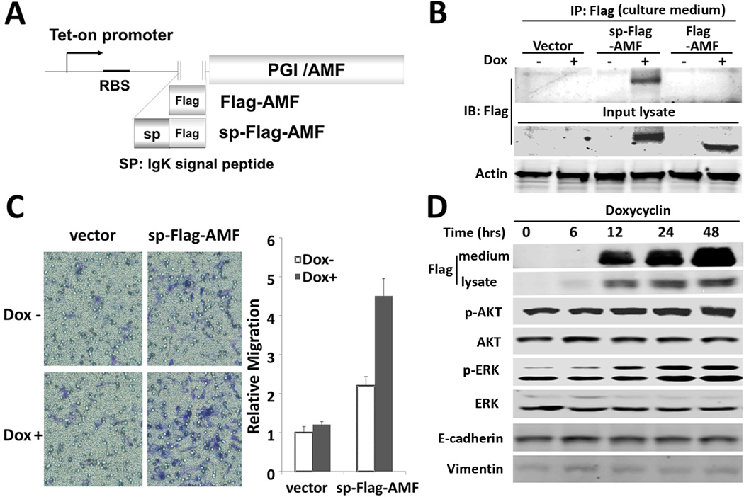
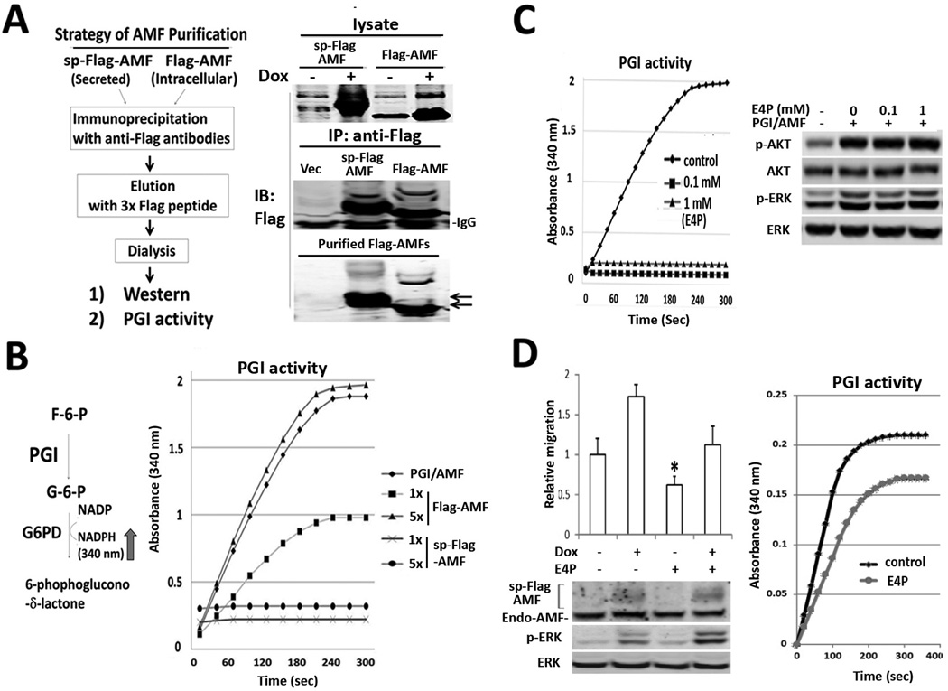
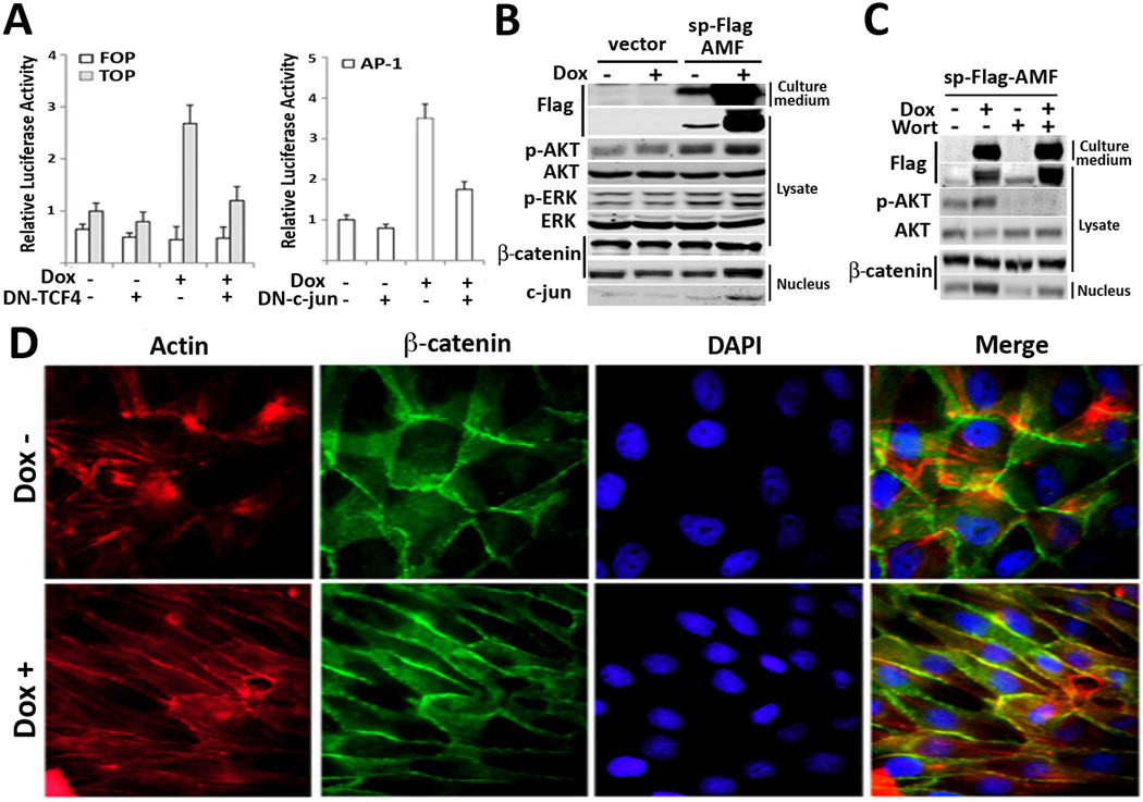
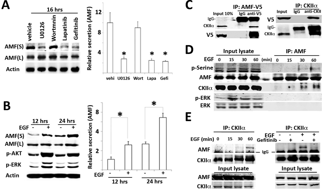
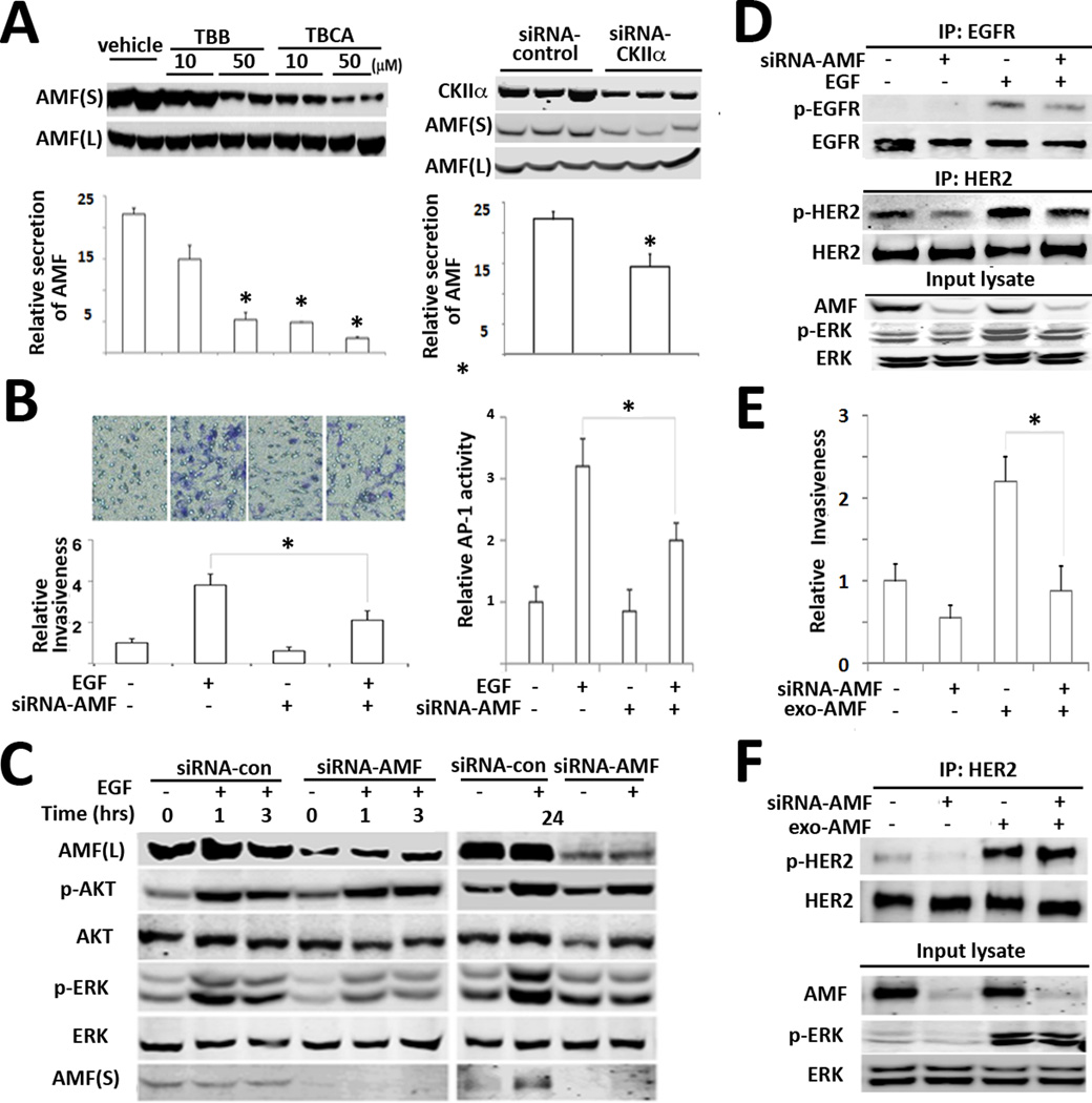
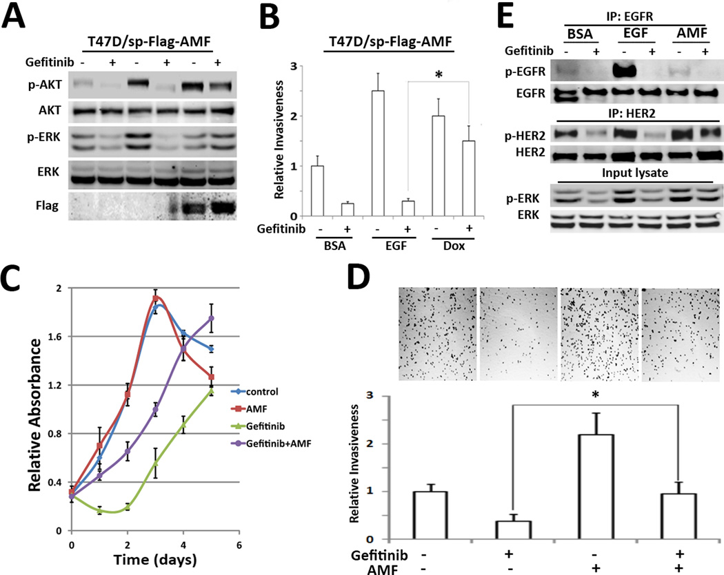
Autocrine motility factor (AMF) enhances invasion by breast cancer cells, but how its secretion and effector signaling are controlled in the tumor microenvironment is not fully understood. In this study, we investigated these issues with a chimeric AMF that is secreted at high levels through a canonical endoplasmic reticulum (ER)/Golgi pathway. Using this tool, we found that AMF enhances tumor cell motility by activating AKT/ERK, altering actin organization, and stimulating β-catenin/TCF and activating protein 1 transcription. EGF enhanced secretion of AMF through its casein kinase II-mediated phosphorylation. RNA interference-mediated attenuation of AMF expression inhibited EGF-induced invasion by suppressing extracellular signal-regulated kinase signaling. Conversely, exogenous AMF overcame the inhibitory effect of EGF receptor inhibitor gefitinib on invasive motility by activating HER2 signaling. Taken together, our findings show how AMF modulates EGF-induced invasion while affecting acquired resistance to cytotoxic drugs in the tumor microenvironment.


Conflict of interest statement

There are no potential conflicts to declare.

Figures

Figure 1
Figure 1
Signal peptide-fused AMF enhances cell motility via activation of PI3K/AKT and MAPK/ERK. A, Illustration of IgK signal peptide (sp) fused to human PGI/AMF. B, Intracellular expression and secretion of sp-Flag-AMF and Flag-AMF were determined in culture medium, after immunoprecipitation (IP) with anti-Flag antibodies were carried out in stable cells which were treated with doxycycline (Dox, 1 εg/ml) for 24 hrs. C, Transwell migration assay in T47D/sp-Flag-AMF cells. Cells were induced with Dox for 24 hrs. and then subjected to migration assay for 24 hrs. Data are shown as mean ± SE of three independent experiments. D, Phosphorylation of AKT and ERK, expression of EMT markers in T47D/sp-Flag-AMF cells which were treated with Dox at the time points indicated. Total cell lysates were separated by SDS-PAGE and immunoblotted.
Figure 2
Figure 2
Enzyme activity of PGI/AMF is not indispensable for AKT/ERK activation. A. Strategy of Flag-AMF purification (left). Products are subjected to immunoblotting analysis during serial steps of purification (right). B, Enzyme activity of glucose-6-phosphate isomerase was analyzed by spectrophotometer, which detects NADPH production by coupling reaction of glucose-6-phosphate dehydrogenase (G6PD) and PGI (left). 2.5 ng (1×) of Flag-AMF and sp-Flag-AMF were measured for their activities (right). C, Effect of E4P on PGI activity and AKT/ERK activation. 2.5 ng of purified PGI/AMF were preincubated with 0, 0.1, 1 mM of E4P at tube, respectively and subjected to enzymatic spectrophotometer assay (left). T47D parental cells were induced with E4P-pretreated PGI/AMF and analyzed (right). D, Effect of E4P on cell motility and intracellular PGI/AMF enzyme activity in T47D/sp-Flag-AMF cells. The cells migrated to bottom wells contained compounds as indicated for 16 hrs. PGI activity of lysate (25 εg) was measured in T47D/sp-Flag-AMF cells treated with 1 mM E4P for 16 hrs.
Figure 3
Figure 3
Chimeric AMF activates β-catenin/TCF and AP-1 transcription, a marker of motility and invasiveness. A, Transcriptional reporter assays in T47D/sp-flag-AMF cells which were transiently transfected with TCF response (TOPFlASH) or AP-1 response (AP-1 Luc) reporters or dominant negative (DN)-TCF and DN-c-jun. Doxycycline was simultaneously treated with transfection. Data are displayed as mean ± SD of three independent experiments. B, T47D/sp-Flag-AMF cells were induced by Dox for 24hrs and then lysates, nuclear fraction were immunoblotted for expression levels of nuclear β-catenin and c-jun. C, T47D/sp-Flag-AMF cells were treated with Wortmannin inhibitor for 24 hrs to analyze its effect on β-catenin translocation. D, Immunofluorescence images of T47D/sp-Flag-AMF cells with Dox induction for 96 hrs. Phalloidin and DAPI were used for Actin and nucleus staining respectively. Lateral extension of cells is shown upon Dox stimulus (original magnification, ×100).
Figure 4
Figure 4
EGF enhances CKII-mediated AMF secretion. A, HER2-overexpressing SKBR3 cells were starved with serum-free medium and treated with inhibitors for 16 hrs. respectively. 100 nM Wortmannin (PI3K inhibitor), 20 εM U0126 (MAPK inhibitor), 10 εM Lapatinib (dual EGFR/HER2 inhibitor), and 10 εM Gefinitib (EGFR specific inhibitor) were used. Culture supernatant (S) was concentrated with 30-kDa cut-off membrane filters. After viable cell counting, culture supernatant is subjected to immunoblotting. S and L indicate culture medium and lysate respectively. B, T47D cells were stimulated with EGF (100 ng/ml) as indicated time points. P-AKT/p-ERK is positive control in response to EGF. Data are representative as the mean ± SE of three independent experiments. * P > 0.05 significantly different from vehicle. C, Lysates were prepared from EBNA293 cells transiently transfected with AMF-V5 and immunoprecipitated with indicated antibodies. IgG indicates heavy chain of IgG. D, T47D cells were stimulated with EGF after serum starvation as indicated time course. IP were performed with anti-AMF antibodies. E. IP with anti-CKII antibodies in T47D cells stimulated with EGF at indicated time points (left). The cells were pretreated with Gefitinib (10 εM) for 2 hours and induced with EGF for 1hr.
Figure 5
Figure 5
AMF knockdown suppresses EGF-induced invasiveness. A, MDA-MB 231 cells were treated with CKII inhibitor (TBB or TBCA) as indicated concentration for 16 hrs. Intracellular and extracellular AMF levels were determined in immunoblot (left). The cells were treated with siRNA-CKIIα and then secreted AMF level was analyzed (right). Bars indicate the mean ± SE of three independent experiments (* p > 0.05). B, after treatment with siRNA-AMF for 48 hrs., T47D cells were counted and loaded into upper side of transwell and chemoattractant of EGF (100 ng/ml) was added at bottom wells (left). Minus (−) indicates vehicle or siRNA-control. AP-1 transcriptional activity was determined in T47D cells, which were pretreated with siRNA-AMF for 24 hrs and then transfected with AP-1 reporter for 24 hrs. (right). Values are expressed as the mean ± SE of three replicates (* p> 0.05). C, Effect of AMF knockdown on EGF-induced p-AKT/p-ERK. After T47D cells were transfected with siRNA-AMF and starved in serum-free medium for 16 hrs. EGF was treated as indicated time points. D, IP with anti-EGFR or HER2 in MDA-MB-231 cells transfected with siRNA-AMF or control siRNA (−) for 48hr and starved, stimulated with EGF for 5 min. E. Rescue experiment of invasion with exogenous AMF in AMF-knockdown MDA-MB-231 cells. The cells were transfected with siRNA-AMF for 48 hrs and subjected to transwell assay. D. AMF-knockdown cells were stimulated with purified AMF and then IP were performed to analyze p-HER2 level.
Figure 6
Figure 6
AMF overexpression overcomes Gefitinib-inhibited invasion. A, Effect of AMF on Gefitinib-pretreated T47D/sp-Flag-AMF cells. After pretreatment with Gefitinib (10 µM) for 2 hrs, Dox and EGF were treated for 24 hrs. P-AKT/p-ERK statuses were analyzed. B, Gefitinib-pretreated cells were subjected to transwell assay with EGF or Dox induction. C, the growth of MDA-MB-231 cells was measured under Gefitinib and AMF treatment, using MTT assay. D, MDA-MB-231 cells were subjected to transwell assay. Each bottom side contained Gefitinib or purified AMF as indicated. Photographs show settle-down cells in bottom wells after cell invasion (* p> 0.05). E. MDA-MB-231cells were pretreated with Gefitinib and then stimulated with EGF or AMF for 5 min respectively. phosphorylation of EGFR and HER2 were analyzed with IP.

References

    1. Watanabe H, Takehana K, Date M, Shinozaki T, Raz A. Tumor cell autocrine motility factor is the neuroleukin/phosphohexose isomerase polypeptide. Cancer Res. 1996;56:2960–2963. - PubMed
    1. Gurney ME. Neuroleukin: basic biology and functional interaction with human immunodeficiency virus. Immunol Rev. 1987;100:203–223. - PubMed
    1. Xu W, Seiter K, Feldman E, Ahmed T, Chiao JW. The differentiation and maturation mediator for human myeloid leukemia cells shares homology with neuroleukin or phosphoglucose isomerase. Blood. 1996;87:4502–4506. - PubMed
    1. Fairbank M, St-Pierre P, Nabi IR. The complex biology of autocrine motility factor/phosphoglucose isomerase (AMF/PGI) and its receptor, the gp78/AMFR E3 ubiquitin ligase. Mol Biosyst. 2009;5:793–801. - PubMed
    1. Bertini R, Howard OM, Dong HF, Oppenheim JJ, Bizzarri C, Sergi R, et al. Thioredoxin, a redox enzyme released in infection and inflammation, is a unique chemoattractant for neutrophils, monocytes, and T cells. J Exp Med. 1999;189:1783–1789. - PMC - PubMed

Publication types

MeSH terms

Substances