

DJ-1 protects against cell death following acute cardiac ischemia-reperfusion injury
- PMID: 24577080
- PMCID: PMC3944257
- DOI: 10.1038/cddis.2014.41
DJ-1 protects against cell death following acute cardiac ischemia-reperfusion injury
Abstract
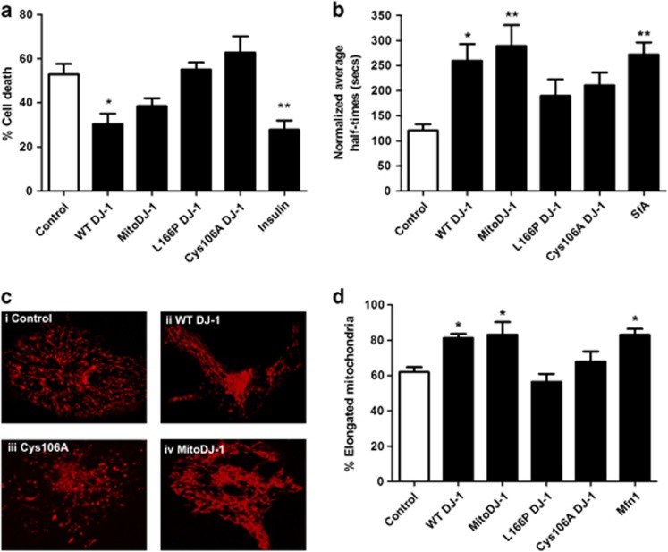
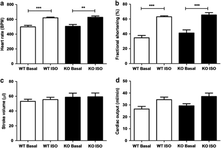
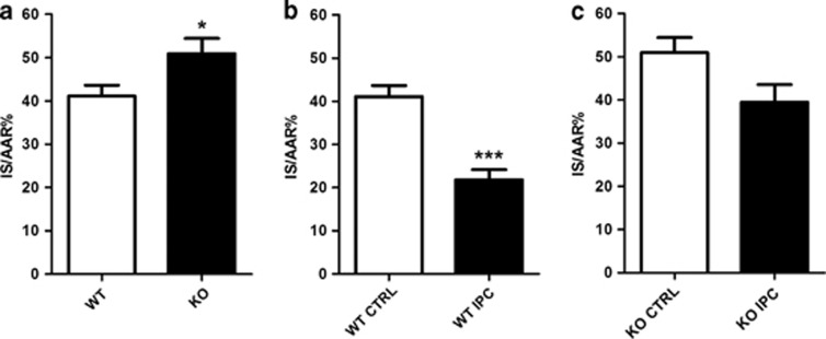
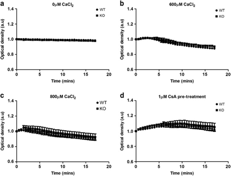
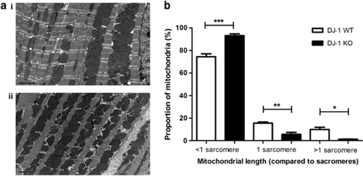
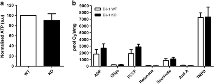
Novel therapeutic targets are required to protect the heart against cell death from acute ischemia-reperfusion injury (IRI). Mutations in the DJ-1 (PARK7) gene in dopaminergic neurons induce mitochondrial dysfunction and a genetic form of Parkinson's disease. Genetic ablation of DJ-1 renders the brain more susceptible to cell death following ischemia-reperfusion in a model of stroke. Although DJ-1 is present in the heart, its role there is currently unclear. We sought to investigate whether mitochondrial DJ-1 may protect the heart against cell death from acute IRI by preventing mitochondrial dysfunction. Overexpression of DJ-1 in HL-1 cardiac cells conferred the following beneficial effects: reduced cell death following simulated IRI (30.4±4.7% with DJ-1 versus 52.9±4.7% in control; n=5, P<0.05); delayed mitochondrial permeability transition pore (MPTP) opening (a critical mediator of cell death) (260±33 s with DJ-1 versus 121±12 s in control; n=6, P<0.05); and induction of mitochondrial elongation (81.3±2.5% with DJ-1 versus 62.0±2.8% in control; n=6 cells, P<0.05). These beneficial effects of DJ-1 were absent in cells expressing the non-functional DJ-1(L166P) and DJ-1(Cys106A) mutants. Adult mice devoid of DJ-1 (KO) were found to be more susceptible to cell death from in vivo IRI with larger myocardial infarct sizes (50.9±3.5% DJ-1 KO versus 41.1±2.5% in DJ-1 WT; n≥7, P<0.05) and resistant to cardioprotection by ischemic preconditioning. DJ-1 KO hearts showed increased mitochondrial fragmentation on electron microscopy, although there were no differences in calcium-induced MPTP opening, mitochondrial respiratory function or myocardial ATP levels. We demonstrate that loss of DJ-1 protects the heart from acute IRI cell death by preventing mitochondrial dysfunction. We propose that DJ-1 may represent a novel therapeutic target for cardioprotection.
Figures
Similar articles
-
The innate immune receptor NLRX1 is a novel required modulator for mPTP opening: implications for cardioprotection.Basic Res Cardiol. 2025 Aug;120(4):707-725. doi: 10.1007/s00395-025-01124-x. Epub 2025 Jun 19. Basic Res Cardiol. 2025. PMID: 40536683 Free PMC article.
-
Akt protects the heart against ischaemia-reperfusion injury by modulating mitochondrial morphology.Thromb Haemost. 2015 Mar;113(3):513-21. doi: 10.1160/TH14-07-0592. Epub 2014 Sep 25. Thromb Haemost. 2015. PMID: 25253080
-
HIF-1 reduces ischaemia-reperfusion injury in the heart by targeting the mitochondrial permeability transition pore.Cardiovasc Res. 2014 Oct 1;104(1):24-36. doi: 10.1093/cvr/cvu172. Epub 2014 Jul 25. Cardiovasc Res. 2014. PMID: 25063991
-
The mitochondrial permeability transition pore and its role in myocardial ischemia reperfusion injury.J Mol Cell Cardiol. 2015 Jan;78:23-34. doi: 10.1016/j.yjmcc.2014.11.005. Epub 2014 Nov 14. J Mol Cell Cardiol. 2015. PMID: 25446182 Review.
-
Role of the MPTP in conditioning the heart - translatability and mechanism.Br J Pharmacol. 2015 Apr;172(8):2074-84. doi: 10.1111/bph.13013. Epub 2015 Jan 8. Br J Pharmacol. 2015. PMID: 25393318 Free PMC article. Review.
Cited by
-
Literature-Based Enrichment Insights into Redox Control of Vascular Biology.Oxid Med Cell Longev. 2019 May 16;2019:1769437. doi: 10.1155/2019/1769437. eCollection 2019. Oxid Med Cell Longev. 2019. PMID: 31223421 Free PMC article. Review.
-
Differential effect of DJ-1/PARK7 on development of natural and induced regulatory T cells.Sci Rep. 2015 Dec 4;5:17723. doi: 10.1038/srep17723. Sci Rep. 2015. PMID: 26634899 Free PMC article.
-
DJ-1-mediated p62 degradation delays intervertebral disc degeneration by inhibiting apoptosis of nucleus pulposus cells.Apoptosis. 2023 Oct;28(9-10):1357-1371. doi: 10.1007/s10495-023-01862-0. Epub 2023 Jun 10. Apoptosis. 2023. PMID: 37300741
-
Pretreatment with Sodium Phenylbutyrate Alleviates Cerebral Ischemia/Reperfusion Injury by Upregulating DJ-1 Protein.Front Neurol. 2017 Jun 9;8:256. doi: 10.3389/fneur.2017.00256. eCollection 2017. Front Neurol. 2017. PMID: 28649223 Free PMC article.
-
DJ-1 administration exerts cardioprotection in a mouse model of acute myocardial infarction.Front Pharmacol. 2022 Sep 23;13:1002755. doi: 10.3389/fphar.2022.1002755. eCollection 2022. Front Pharmacol. 2022. PMID: 36210822 Free PMC article.
References
-
- Bonifati V, Rizzu P, van Baren MJ, Schaap O, Breedveld GJ, Krieger E, et al. Mutations in the DJ-1 gene associated with autosomal recessive early-onset parkinsonism. Science. 2003;299:256–259. - PubMed
-
- Zhang L, Shimoji M, Thomas B, Moore DJ, YuSW, Marupudi NI, et al. Mitochondrial localization of the Parkinson's disease related protein DJ-1: implications for pathogenesis. Hum Mol Genet. 2005;14:2063–2073. - PubMed
-
- Kinumi T, Kimata J, Taira T, Ariga H, Niki E. Cysteine-106 of DJ-1 is the most sensitive cysteine residue to hydrogen peroxide-mediated oxidation in vivo in human umbilical vein endothelial cells. Biochem Biophys Res Commun. 2004;317:722–728. - PubMed
Publication types
MeSH terms
Substances
Grants and funding
LinkOut - more resources
Full Text Sources
Other Literature Sources
Medical
Molecular Biology Databases
Research Materials
Miscellaneous
