. 2014 Feb 27;5(2):e1091.
doi: 10.1038/cddis.2014.62.

Impact of human papilloma virus infection on the response of head and neck cancers to anti-epidermal growth factor receptor antibody therapy

Affiliations

Impact of human papilloma virus infection on the response of head and neck cancers to anti-epidermal growth factor receptor antibody therapy

M Pogorzelski et al. Cell Death Dis. .

Abstract

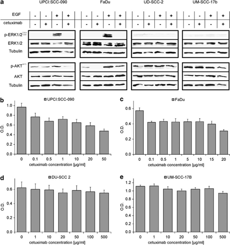
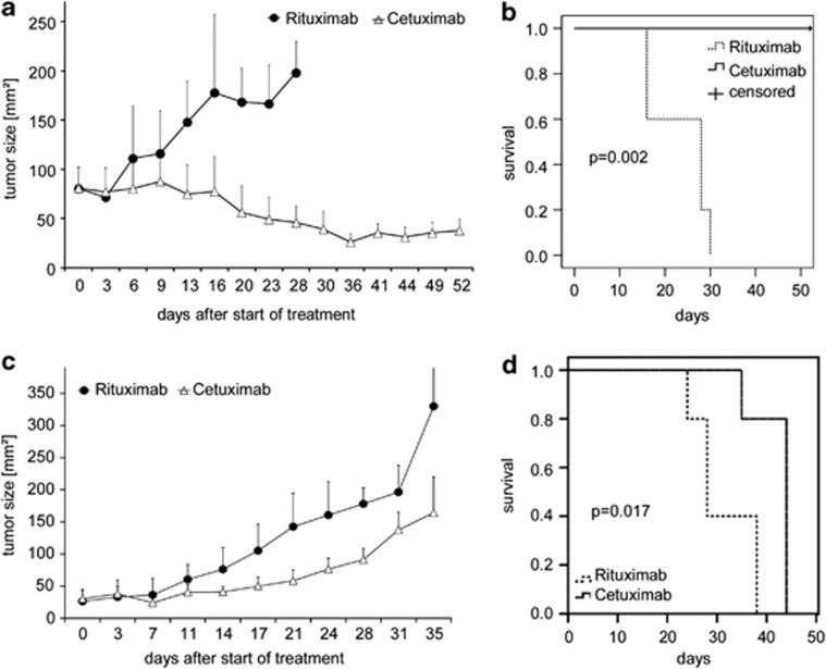
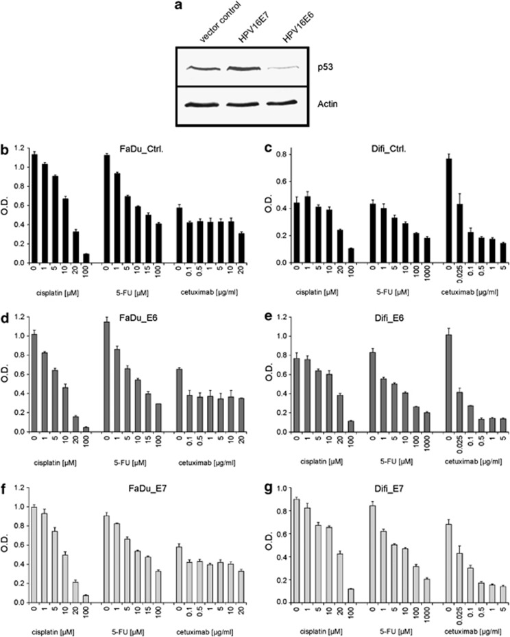
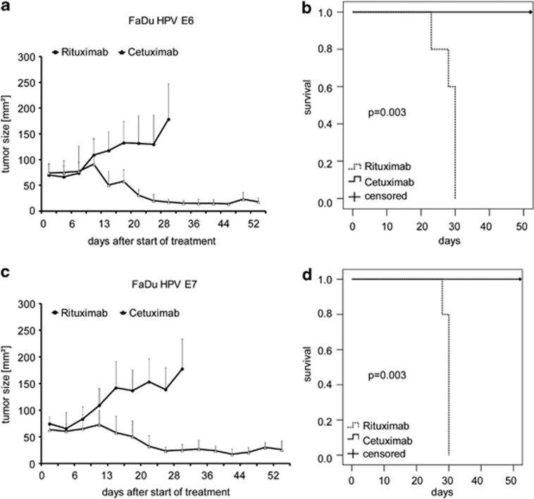
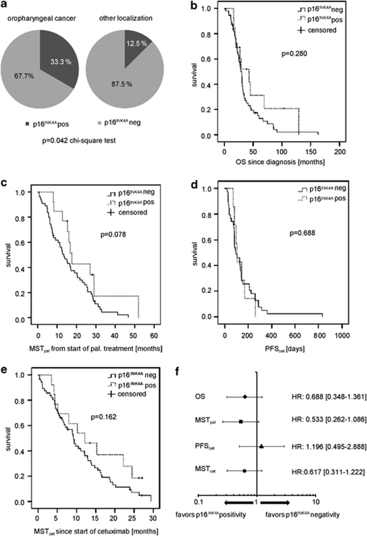
Infection with human papillomaviruses (HPVs) characterizes a distinct subset of head and neck squamous cell cancers (HNSCCs). HPV-positive HNSCC preferentially affect the oropharynx and tonsils. Localized HPV-positive HNSCCs have a favorable prognosis and treatment outcome. However, the impact of HPV in advanced or metastatic HNSCC remains to be defined. In particular, it is unclear whether HPV modulates the response to cetuximab, an antibody targeting the epidermal growth factor receptor (EGFR), which is a mainstay of treatment of advanced HNSCC. To this end, we have examined the sensitivity of HPV-positive and -negative HNSCC models to cetuximab and cytotoxic drugs in vitro and in vivo. In addition, we have stably expressed the HPV oncogenes E6 and E7 in cetuximab-sensitive cancer cell lines to specifically investigate their role in the antibody response. The endogenous HPV status or the expression of HPV oncogenes had no significant impact on cetuximab-mediated suppression of EGFR signaling and proliferation in vitro. Cetuximab effectively inhibited the growth of E6- and E7-expressing tumors grafted in NOD/SCID mice. In support, formalin-fixed, paraffin-embedded tumor samples from cetuximab-treated patients with recurrent or metastatic HNSCC were probed for p16(INK4a) expression, an established biomarker of HPV infection. Response rates (45.5% versus 45.5%) and median progression-free survival (97 versus 92 days) following cetuximab-based therapy were similar in patients with p16(INK4A)-positive and p16(INK4A)-negative tumors. In conclusion, HPV oncogenes do not modulate the anti-EGFR antibody response in HSNCC. Cetuximab treatment should be administered independently of HPV status.


Figures

Figure 1
Figure 1
Sensitivity of EGFR-positive cancer cell lines to the anti-EGFR antibody cetuximab in relation to HPV status. (a) Inhibition of constitutive and ligand-induced (EGF 10 ng/ml for 10 min) phosphorylation of ERK and AKT by cetuximab (1 μg/ml for 2 h) in HPV-positive UPCI:SCC-090 and HPV-negative FaDu cells. Note that ERK and AKT phosphorylation are unresponsive to EGF or cetuximab in UD-SCC-2 (HPV positive) and UM-SCC-17b (HPV-negative) cells. (be) The HPV-positive UPCI:SCC-090 and UD-SCC-2 and the HPV-negative FaDu and UM-SCC-17b HNSCC cell lines were grown in the presence of cetuximab at the indicated concentrations for 72 h (FaDu) or 96 h (UPCI:SCC-090, UD-SCC-2 and UM-SCC-17b). Optical density (OD; +s.d.) of formazan solution from three independent 3-[4,5–dimethylthiazol–2–yl]-2,5-diphenyl tetrazolium bromide assays is shown
Figure 2
Figure 2
Effective treatment of established HPV-negative and HPV-positive tumors by treatment with cetuximab in vivo. Palpable flank tumors were established by subcutaneous injection of (a and b) FaDu (HPV-negative) or (c and d) UPCI:SCC-090 (HPV-positive) cells in NOD/SCID mice. After 14 days, tumor-bearing mice were treated twice weekly by intraperitoneal injections of cetuximab (1 mg, white triangles) or the control antibody rituximab (1 mg, black circles). (a and c) Tumor growth was monitored by palpation and quantified using a caliper. Mean bidimensional tumor sizes (+s.d.) of a representative experiment (five mice per treatment group) are given. (b and d) Kaplan–Meier plots of survival of tumor-bearing NOD/SCID mice. (b) HPV-negative FaDu; (d) HPV-positive UPCI:SCC-090. Mice were treated as in a and c with cetuximab (solid line), or the control antibody rituximab (dashed line). Cetuximab-treated mice exhibited a significantly prolonged survival as compared with rituximab-treated mice. In the FaDu model, median survival time for cetuximab-treated mice was not reached. It was 28 days for mice receiving rituximab (P=0.002, log rank test). In the UPCI:SCC-090 model, median survival time for cetuximab-treated mice was 44 days, and 28 days for rituximab-treated mice (P=0.017, log rank test)
Figure 3
Figure 3
The HPV oncogenes E6 and E7 do not modulate the cetuximab response of EGFR-positive cancer cells in vitro. The EGFR-positive cetuximab-sensitive cancer cell lines FaDu, Difi and A431 were retrovirally transduced to stably express the HPV16 oncogenes E6 and E7 or a control vector. (a) Increased and decreased p53 levels in response to expression of HPV16 E7 or E6. A representative immunoblot of FaDu cells is shown; similar results were obtained in Difi and A431 (not shown). (bg) FaDu and Difi cells expressing HPV16 E6, E7 or a control vector (ctrl) were grown for 72 h in the presence of cetuximab, cisplatin or 5-fluorouracil (5-FU) at the indicated concentrations. Optical density (OD; +s.d.) of formazan solution from three independent 3-[4,5–dimethylthiazol–2–yl]-2,5-diphenyl tetrazolium bromide assays is shown
Figure 4
Figure 4
Impact of enforced HPV E6 and E7 expression on the cetuximab response of HPV-negative HNSCC cells in vivo. (a and c) Tumor growth following injection of FaDu cells stably expressing HPV16 E6 (a) or HPV16 E7 oncogenes (c) in NOD/SCID mice. After 14 days, tumor-bearing mice received biweekly intraperitoneal injections of cetuximab (1 mg, white triangles) or the control antibody rituximab (black circles). Mean bidimensional tumor sizes (+s.d.) of five mice per group are given. Rapid and sustained tumor shrinkage was observed following cetuximab treatment. In contrast, tumors from rituximab-treated mice continuously progressed. (b and d) Kaplan–Meier plots of survival of NOD/SCID mice bearing FaDu tumors expressing HPV16 E6 (b) or HPV16 E7 (d). Mice were treated with cetuximab (solid line), or the control antibody rituximab (dashed line) as in a and c. Cetuximab-treated mice showed significantly prolonged survival as compared with rituximab-treated mice. Median survival time was not reached for cetuximab-treated FaDu HPV E6 tumour-bearing mice. It was 30 days for mice treated with rituximab (P=0.003, log rank test). In the FaDu HPV E7 model, median survival time for cetuximab-treated mice was not reached, and was 30 days for rituximab-treated mice (P=0.003, log rank test)
Figure 5
Figure 5
Clinical outcome and cetuximab response of patients with recurrent or metastatic HNSCC in relation to p16INK4A expression. (a) Immunohistochemically detectable p16INK4A expression in relation to HNSCC localization. Oropharyngeal cancers exhibited a statistically significant higher prevalence of p16INK4A positivity as compared with HNSCC of other localizations. Patients with tumors expressing p16INK4A were considered HPV positive. (b) Kaplan–Meier plot of overall survival (OS) from first diagnosis of patients with p16INK4A-negative (solid line) and p16INK4A-positive (dashed line) recurrent or metastatic HNSCC. Patients with p16INK4A-positive tumors demonstrated a numerically prolonged OS (42.97 versus 30.03 months), which did not reach statistical significance (P=0.280, log rank). (c) Kaplan–Meier plot of median survival time from initiation of palliative treatment (MSTpal) in patients with p16INK4A-negative (solid line) and p16INK4A-positive (dashed line) relapsed or metastatic HNSCC. Patients with p16INK4A-positive tumors had a numerically prolonged MSTpal (17.64 versus 13.44 months). The difference did not reach statistical significance (P=0.078, log rank). (d) Kaplan–Meier plot of median progression-free survival following cetuximab treatment (PFScet) in patients with p16INK4A-negative (solid line) and p16INK4A-positive (dashed line) HNSCC. Median PFScet was identical in both groups (P=0.688, log rank). (e) Kaplan–Meier plot of median survival time from start of cetuximab treatment (MSTcet) in patients with p16INK4A-negative (solid line) and p16INK4A-positive (dashed line) recurrent or metastatic HNSCC. Patients with p16INK4A-positive HNSCC showed a trend toward longer MSTcet as compared with patients with p16INK4A-negative HNSCC (12.16 versus 9.23 months, P=0.162, log rank). (f) Forrest blot of hazard ratios (HR) and 95% confidence intervals (CI) of OS, MSTpal, PFScet and MSTcet. A HR<1 favors p16INK4A positivity, and a HR>1 favors p16INK4A negativity. There was no trend toward an interaction between the PFScet and p16INK4A status

Similar articles

Cited by

References

    1. Leemans CR, Braakhuis BJ, Brakenhoff RH. The molecular biology of head and neck cancer. Nat Rev Cancer. 2011;11:9–22. - PubMed
    1. Chaturvedi AK, Engels EA, Pfeiffer RM, Hernandez BY, Xiao W, Kim E, et al. Human papillomavirus and rising oropharyngeal cancer incidence in the United States. J Clin Oncol. 2011;29:4294–4301. - PMC - PubMed
    1. Hong AM, Grulich AE, Jones D, Lee CS, Garland SM, Dobbins TA, et al. Squamous cell carcinoma of the oropharynx in Australian males induced by human papillomavirus vaccine targets. Vaccine. 2010;28:3269–3272. - PubMed
    1. Mork J, Lie AK, Glattre E, Hallmans G, Jellum E, Koskela P, et al. Human papillomavirus infection as a risk factor for squamous-cell carcinoma of the head and neck. N Engl J Med. 2001;344:1125–1131. - PubMed
    1. Nasman A, Attner P, Hammarstedt L, Du J, Eriksson M, Giraud G, et al. Incidence of human papillomavirus (HPV) positive tonsillar carcinoma in Stockholm, Sweden: an epidemic of viral-induced carcinoma. Int J Cancer. 2009;125:362–366. - PubMed

Publication types

MeSH terms

Substances