. 2014 Feb 27;5(2):e1095.
doi: 10.1038/cddis.2014.47.

MicroRNA-21 regulates T-cell apoptosis by directly targeting the tumor suppressor gene Tipe2

Affiliations

MicroRNA-21 regulates T-cell apoptosis by directly targeting the tumor suppressor gene Tipe2

Q Ruan et al. Cell Death Dis. .

Abstract

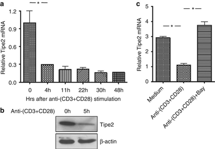
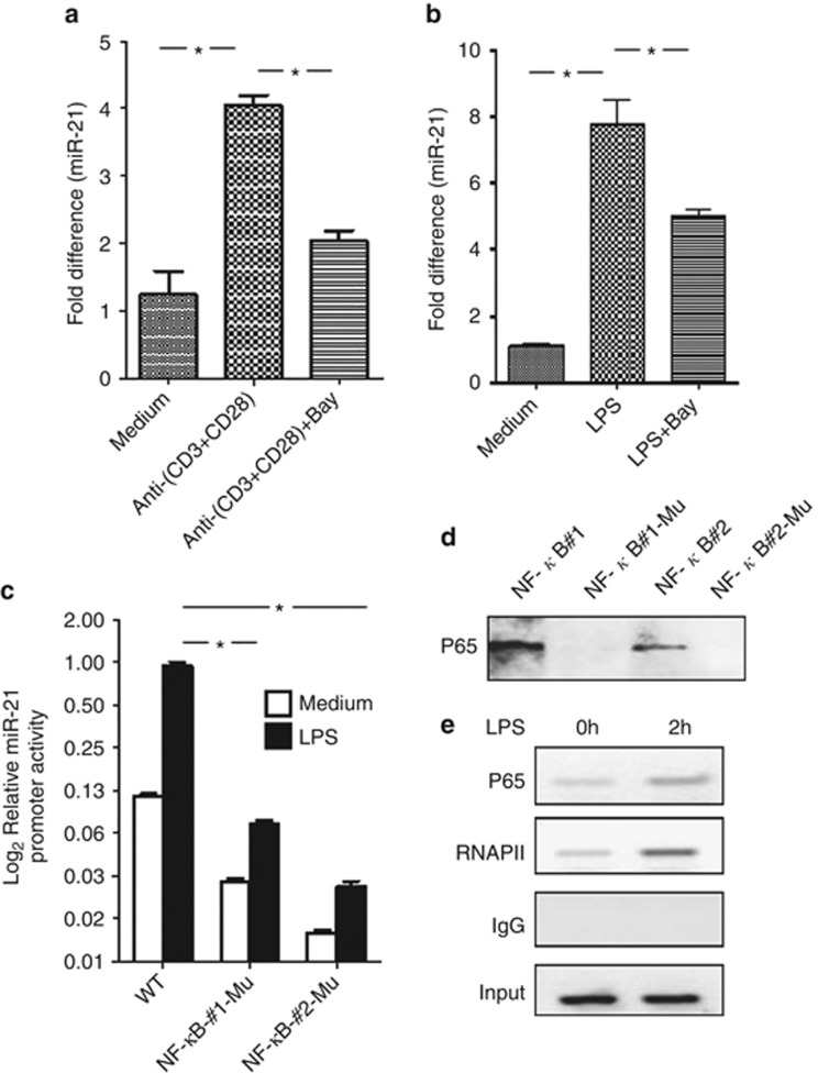
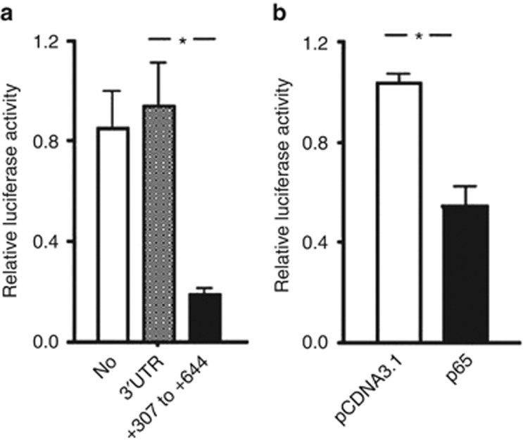
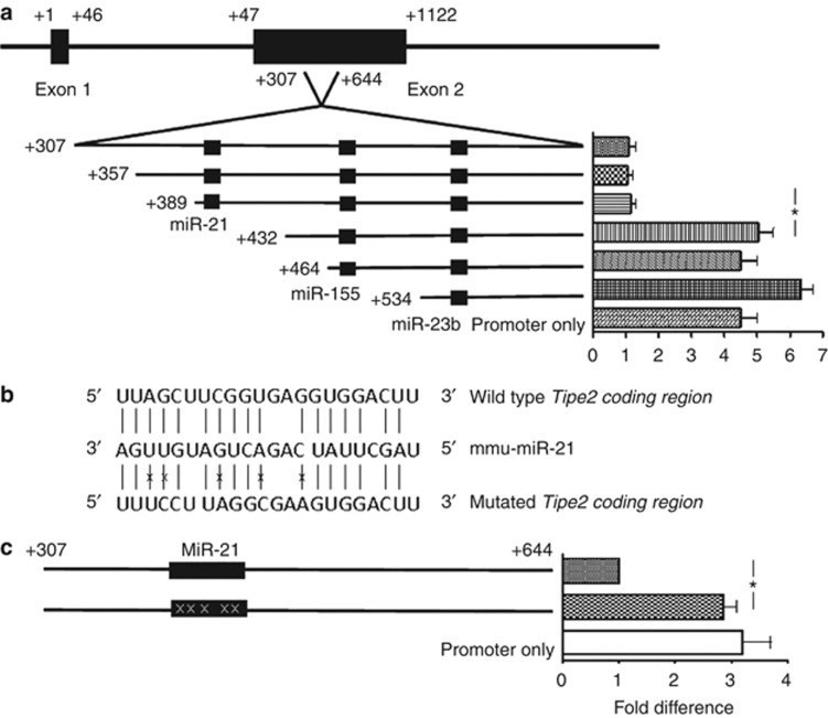
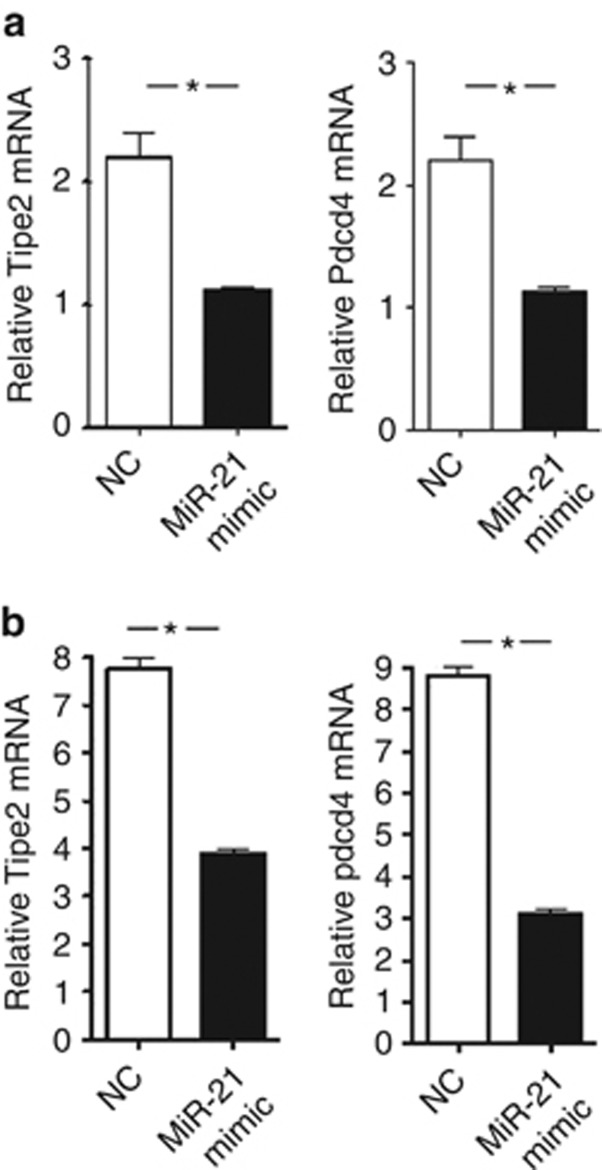
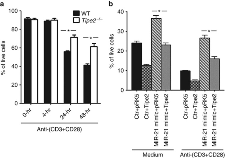
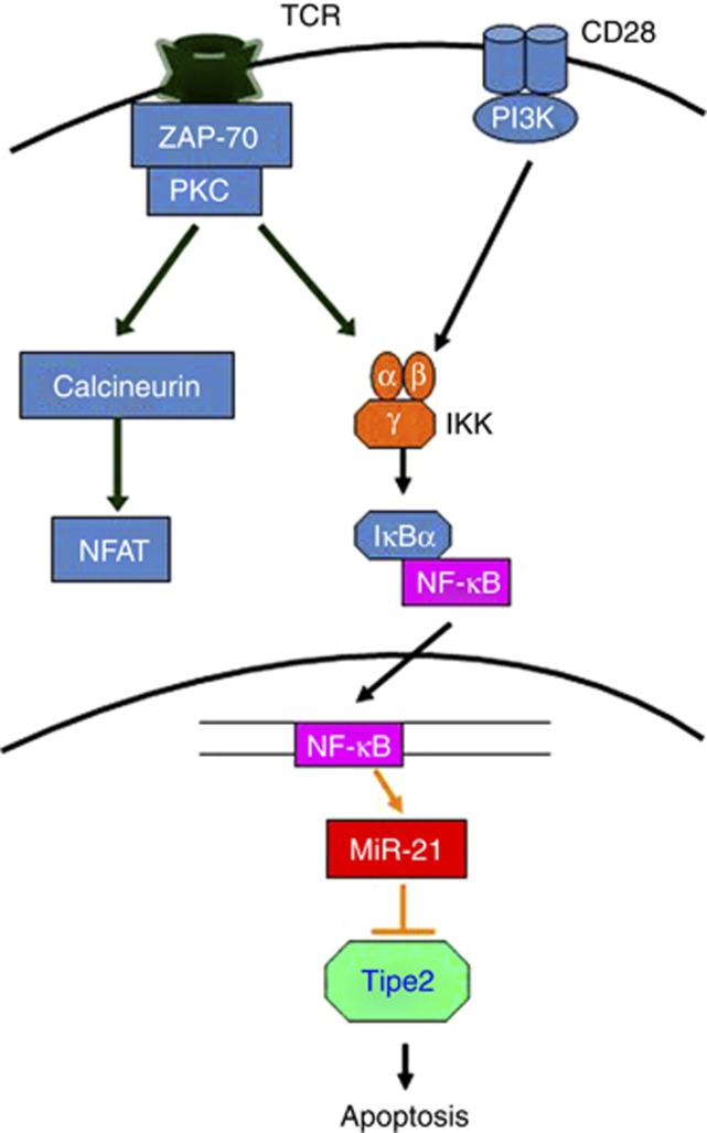
MicroRNAs (MiRs) are short noncoding RNAs that can regulate gene expression. It has been reported that miR-21 suppresses apoptosis in activated T cells, but the molecular mechanism remains undefined. Tumor suppressor Tipe2 (or tumor necrosis factor-α-induced protein 8 (TNFAIP8)-like 2 (TNFAIP8L2)) is a newly identified anti-inflammatory protein of the TNFAIP8 family that is essential for maintaining immune homeostasis. We report here that miR-21 is a direct target of nuclear factor-κB and could regulate Tipe2 expression in a Tipe2 coding region-dependent manner. In activated T cells and macrophages, Tipe2 expression was markedly downregulated, whereas miR-21 expression was upregulated. Importantly, Tipe2-deficient T cells were significantly less sensitive to apoptosis. Conversely, overexpression of Tipe2 in EL-4 T cells increased their susceptibility to activation-induced apoptosis. Therefore, Tipe2 provides a molecular bridge between miR-21 and cell apoptosis; miR-21 suppresses apoptosis in activated T cells at least in part through directly targeting tumor suppressor gene Tipe2.


Figures

Figure 1
Figure 1
NF-κB-dependent downregulation of Tipe2 in T cells and myeloid cells. (a) Murine naive CD4+ T cells were either untreated or treated with plate-bound anti-CD3 (1 μg/ml) and soluble anti-CD28 (1 μg/ml) for the indicated times, and Tipe2 mRNA levels were determined by quantitative PCR. (b) Murine naive CD4+ T cells were either untreated or treated with plate-bound anti-CD3 (1 μg/ml) and soluble anti-CD28 (1 μg/ml) for 5 h, Tipe2 protein levels were determined by western blot analysis. (c) Murine naive CD4+ T cells were treated as in (a) for 4 h in the absence or presence of NF-κB inhibitor Bay 11-7082. Tipe2 mRNA levels were determined by quantitative PCR. *P<0.02. The results are representative of three independent experiments
Figure 2
Figure 2
NF-κB binds to and activates the miR-21 promoter. (a) Murine naive CD4+ T cells were either untreated or treated with plate-bound anti-CD3 (1 μg/ml) and soluble anti-CD28 (1 μg/ml) for 6 h in the absence or presence of NF-κB inhibitor Bay 117-82, and miR-21 levels were determined by quantitative PCR. *P<0.01. (b) Murine bone marrow-derived macrophages were either untreated or treated with LPS (100 ng/ml) for 4 h in the absence or presence of NF-κB inhibitor Bay 117-82. MiR-21 levels were determined by quantitative PCR. *P<0.02. (c) RAW264.7 cells were transiently transfected with WT or NF-κB site-mutated murine miR-21 promoter luciferase construct. After 24 h, cells were either untreated or treated with LPS (100 ng/ml) for 6 h, and the luciferase activities measured. The promoter activity is presented as fold increase over untreated cells transfected with NF-κB site no. 2 mutated construct. To normalize the transfection efficiency across samples, the Renilla luciferase expression vector pRL-TK was included as an internal control. *P<0.01. (d) Nuclear extracts were prepared from mouse bone marrow-derived macrophages after stimulation for 2 h with LPS. Biotinylated miR-21 oligonucleotides containing NF-κB binding site nos. 1 or 2 or their mutants were absorbed by streptavidin–agarose beads, and then added to the nuclear extracts. The amount of p65 proteins in the precipitates were assessed by immunoblotting with anti-p65. (e) Mouse bone marrow-derived macrophages were treated with LPS (100 ng/ml). After 2 h, cells were fixed, and ChIP was performed using anti-p65, anti-RNA polymerase II (PolII), or control IgG. Immunoprecipitated DNA was analyzed by PCR using primers for NF-κB site on miR-21 promoter. Data are representative of two independent experiments
Figure 3
Figure 3
Tipe2 coding region, but not 3′-UTR, is responsible for the downregulation of Tipe2. EL4 cells were transiently transfected with murine Tipe2 promoter luciferase construct with Tipe2 3′-UTR (A) or coding region (+307 to +644) (a and b) inserted downstream of the luciferase gene but upstream of polyA signal sequence. For (b), expression vector for full-length p65, or the empty vector as indicated, was co-transfected. At 24 h after transfection, cells were treated with phorbol myristyl acetate (PMA) (50 ng/ml) and ionomycin (1 μM) for 5 h, and the luciferase activities measured. The promoter activity is presented as fold increase over cells transfected with murine miR-21 promoter only luciferase construct (a) or empty vector (b). To normalize the transfection efficiency across samples, the Renilla luciferase expression vector pRLTK was included as an internal control. *P<0.01. Data are representative of three independent experiments
Figure 4
Figure 4
MiR-21 targets Tipe2 in a coding region-dependent manner. (a) Deletion mutants of Tipe2 coding region (+307 to +644) were analyzed in the luciferase reporter assay. Putative binding site for miR-21, miR-155 and miR-23b are indicated. (b) The predicted miR-21 binding site on murine Tipe2 coding region and the mutation strategy is shown. The ‘X' indicates the mutated sites. (c) WT and miR-21 binding site-mutated Tipe2 coding region constructs were analyzed in the luciferase reporter assay. The ‘X' indicates the mutated sites. *P<0.02. Data are representative of three independent experiments
Figure 5
Figure 5
MiR-21 regulates endogenous Tipe2 expression. RAW264.7 (a) and EL4 (b) cells were transfected with miR-21 mimic or negative control as indicated. At 24 h after transfection, miR-21, Tipe2 and Pdcd4 mRNA levels were determined by real-time RT-PCR. *P<0.02. Data are representative of three independent experiments
Figure 6
Figure 6
Tipe2 deficiency in T cells renders them resistant to death and miR-21 regulates T-cell apoptosis through Tipe2. (a) Splenic naive CD4+ T cells were isolated from WT and Tipe2−/− mice (n=3) and either untreated or treated with plate-bound anti-CD3 (1 μg/ml) and soluble anti-CD28 (1 μg/ml) for the indicated times. Cells were then stained with annexin V and 7AAD, and the degree of apoptosis was analyzed by flow cytometry. (b) EL4 cells were transfected with miR-21 mimic or negative control together with Tipe2-overexpressing construct or an empty vector. After 24 h, cells were either untreated or treated with plate-bound anti-CD3 (1 μg/ml) and anti-CD28 (1 μg/ml). After 48 h, cells were stained with annexin V and 7AAD, and the degree of apoptosis was analyzed by flow cytometry. *P<0.01. Data are representative of three independent experiments
Figure 7
Figure 7
NF-κB inhibits activation-induced T-cell apoptosis through the miR-21–Tipe2 axis. TCR and anti-CD28 stimulation activate NF-κB through IKK (by degrading the inhibitor protein IκB). After activation, NF-κB translocates into the nucleus and turns on its target genes, including miR-21. MiR-21 in turn degrades Tipe2 and inhibits activation-induced T-cell apoptosis

Similar articles

Cited by

References

    1. Bernstein E, Kim SY, Carmell MA, Murchison EP, Alcorn H, Li MZ, et al. Dicer is essential for mouse development. Nat Genet. 2003;35:215–217. - PubMed
    1. Calin GA, Croce CM. MicroRNA signatures in human cancers. Nat Rev Cancer. 2006;6:857–866. - PubMed
    1. He L, Hannon GJ. MicroRNAs: small RNAs with a big role in gene regulation. Nat Rev Genet. 2004;5:522–531. - PubMed
    1. Muljo SA, Ansel KM, Kanellopoulou C, Livingston DM, Rao A, Rajewsky K. Aberrant T cell differentiation in the absence of Dicer. J Exp Med. 2005;202:261–269. - PMC - PubMed
    1. Sonkoly E, Stahle M, Pivarcsi A. MicroRNAs and immunity: novel players in the regulation of normal immune function and inflammation. Semin Cancer Biol. 2008;18:131–140. - PubMed

Publication types

MeSH terms

LinkOut - more resources