. 2014 Sep;74(9):918-33.
doi: 10.1002/dneu.22174. Epub 2014 Mar 27.

Activation of PI3K and R-Ras signaling promotes the extension of sensory axons on inhibitory chondroitin sulfate proteoglycans

Affiliations

Activation of PI3K and R-Ras signaling promotes the extension of sensory axons on inhibitory chondroitin sulfate proteoglycans

Lee Silver et al. Dev Neurobiol. 2014 Sep.

Abstract

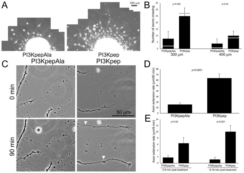
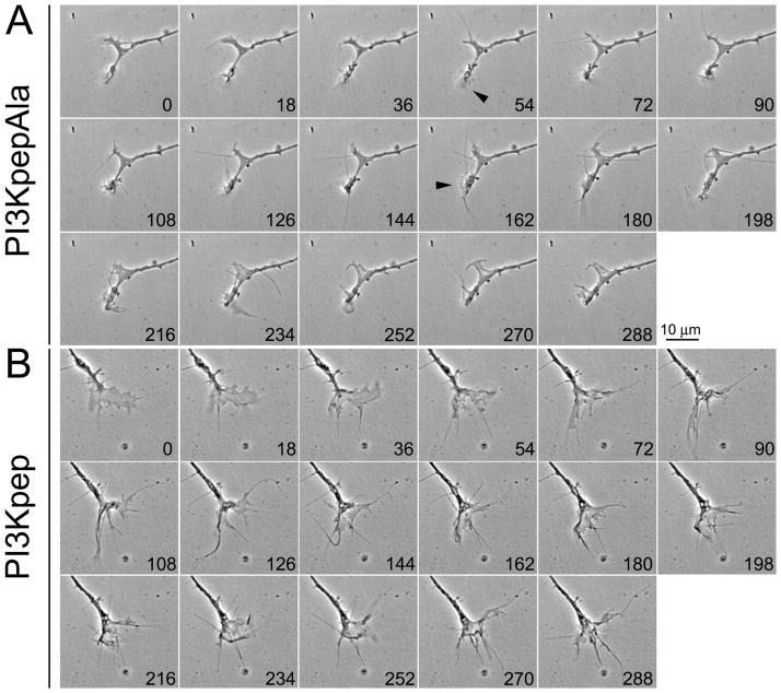
Chondroitin sulfate proteoglycans (CSPGs) are extracellular inhibitors of axon extension and plasticity, and cause growth cones to exhibit dystrophic behaviors. Phosphoinositide 3-kinase (PI3K) is a lipid kinase activated by axon growth promoting signals. In this study, we used embryonic chicken dorsal root ganglion neurons to determine if CSPGs impair signaling through PI3K. We report that CSPGs inhibit PI3K signaling in axons and growth cones, as evidenced by decreased levels of phosphorylated downstream kinases (Akt and S6). Direct activation of PI3K signaling, using a cell permeable phosphopeptide (PI3Kpep), countered the effects of CSPGs on growth cones and axon extension. Both overnight and acute treatment with PI3Kpep promoted axon extension on CSPG-coated substrates. The R-Ras GTPase is an upstream positive regulator of PI3K signaling. Expression of constitutively active R-Ras promoted axon extension and growth cone elaboration on CSPGs and permissive substrata. In contrast, an N-terminus-deleted constitutively active R-Ras, deficient in PI3K activation, promoted axon extension but not growth cone elaboration on CSPGs and permissive substrata. These data indicate that activation of R-Ras-PI3K signaling may be a viable approach for manipulating axon extension on CSPGs.

Keywords: Akt; S6; growth cone; neurite; regeneration.


Figures

FIGURE 1
FIGURE 1
CSPGs decrease the levels of axonal pAkt and pS6. Representative examples of axons and growth cones stained with anti-pAkt (A) and anti-pS6 (B), and counterstained with rhodamine phalloidin to reveal actin filaments. Neurons were cultured in the absence of NGF and then treated with 40 ng/mL NGF or control vehicle for 60 min. NGF treatment elevates the level of staining in axons and growth cones. (C) Quantification of the levels of pAkt and pS6 staining levels in distal axons. For each antibody, data are shown for +/− NGF treatment (E7) and +/− CSPG substrata (E14) in the presence of NGF. n=49–68 axons were sampled per group from 3–4 independent cultures.
FIGURE 2
FIGURE 2
PI3Kpep increases the levels of axonal pAkt and pS6 on CSPG substrata. E14 sensory explants were cultured in the presence of NGF on substrata +/− CSPGs are then treated with PI3Kpep or the control PI3KpepAla for 1 hr. The levels of pAkt and pS6 were quantified in axons as in Figure 1. n=42–49 axons were sampled per group from 3–4 independent cultures.
FIGURE 3
FIGURE 3
PI3Kpep restores growth cone morphology on CSPG substrata and promotes axon crossing onto CSPG substrata. (A) Examples of E14 axons and growth cones stained with anti-α-tubulin and rhodamine phalloidin to reveal actin filaments. On CSPG substrata axons exhibited simplified growth cones with fewer filopodia and less spreading/lamellipodia formation. Treatment with PI3Kpep, relative to PI3KpepAla, for 1 hr restored growth cones on CSPGs to morphologies similar to those observed on control substrata. (B) Quantification of growth cone morphologies as a function of substratum and PI3Kpep and PI3KpepAla treatment. n>43 axons per group, 3–4 cultures per group. Growth cone areas are shown in blue, and filopodia number in red, both as percent of control). (C) Example of axons encountering borders between permissive and CSPG substrata in the presence of PI3KpepAla or PI3Kpep. Axons were stained with anti-α-tubulin and CSPGs were stained as described in Ketschek et al (2012). In the presence of PI3KpepAla axons did not extend onto the CSPG-coated substratum, and instead straddled the border between the substrata. In contrast, in the presence of PI3Kpep axons often crossed onto the CSPG coated substratum. (D) Quantification of the ratio of axons crossing onto the CSPG-coated substratum relative to those that did not, and straddled the border. χ2 analysis on proportion of axons crossing. n=6 explants per group.
FIGURE 4
FIGURE 4
PI3Kpep promotes axon extension on CSPG substrata. (A) 20x composites of axons extending from dorsal root ganglion explants on CSPG-coated substrata overnight in the continuous presence of PI3Kpep or PI3KpepAla. In the presence of PI3Kpep explants exhibited greater densities of axons and the axons extended longer distances than in the presence of PI3KpepAla. (B) Quantification of the number of axons crossing lines at 300 and 400 μm from the base of explants in the representative experiments depicted in panel (A). n=8 explants per group. (C) Examples of axons and growth cones, cultured in NGF and extending on a CSPG-coated substratum, from time-lapse sequences commenced upon treatment with PI3Kpep or PI3KpepAla. The white arrowheads in the 90 min panels denote the position of the growth cone (extent of phase dark axon shaft) at 0 min. (D) Measurement of the distance axons extended during a 90 min period in the experiments depicted in panel (C). PI3Kpep treated axons extended approximately 3 fold faster than those treated with PI3KpepAla. n=29 and 18 axons for PI3KpepAla and PI3Kpep groups from 3 and 4 cultures, respectively. (E) Measurement of the distance axon extended during the first 0–9 and 9–18 min following treatment with PI3KpepAla or PI3Kpep. Dunn multiple comparisons test.
FIGURE 5
FIGURE 5
PI3Kpep treatment promotes growth cone dynamics on CSPG substrata. (A) Time-lapse stills (seconds denoted in panels) of a representative growth cone on a CSPG substratum treated with PI3KpepAla. Note that although lamellipodia formed (e.g., arrowheads at 54 and 162 sec) they were transient and readily retracted into the growth cone. (B) Example of a growth cone treated with PI3Kpep. The panels follow the same format as in (A). In comparison to (A), note the persistence of lamellipodia and that the growth cone exhibits lamellipodia throughout most of the sequence.
FIGURE 6
FIGURE 6
Effects of R-Ras and ΔN-R-Ras ectopic expression on the length and morphology of sensory neuron axons on laminin substrata in the absence of NGF. (A) Examples of the pattern of anti-R-Ras staining in cultured E7 sensory neurons. R-Ras (green) was evident in cell bodies and along axons. (B) Examples of R-Ras staining in distal axons. The R-Ras signal was punctate and present throughout the axon shaft, central and peripheral domains of growth cones. R-Ras was present in both lamellipodia and filopodia. (C) Western analysis of pAkt phosphorylation in serum starved NIH 3T3 cells expressing R-Ras constructs. As a positive control, a 30 treatment with bovine calf serum elevated pAkt levels in cells transfected with the empty vector (vector), relative to serum starved cells. Expression of full length R-Ras in the absence of serum treatment also increased pAkt levels. Expression of the N-terminus (N-term) of R-Ras did not appreciably affect pAkt levels. Expression of ΔN-R-Ras slightly elevated pAkt levels relative to vehicle controls not treated with serum, but to a much lower extent than full length R-Ras. Blot is representative of 3 independent experiments. Quantitative analysis of pAkt/Akt levels, relative to serum starved control conditions, is shown in the bar graph adjacent to the blot. A value of 1 on the Y-axis is reflective of baseline control levels. (D) Expression of R-Ras and ΔN-R-Ras affects growth cone morphology. R-Ras expression resulted in a greater percentage of growth cones with spread lamellipodial morphologies, while ΔN-R-Ras expression increased the percentage of growth cones exhibiting filopodia (p values for comparisons to GFP controls). The distribution of growth cone morphologies also differed between R-Ras and ΔN-R-Ras (p<0.001). (E) Expression of R-Ras and ΔN-R-Ras increased the length of sensory neuron axons. The lengths of axons expression R-Ras and ΔN-R-Ras did not differ.
Figure 7
Figure 7
Effects of R-Ras and ΔN-R-Ras expression on the morphology of growth cones and the length of sensory neuron axons on CSPG substrata. A concentration of 5 ng/mL NGF was present in all experimental groups to provide neurotrophic support. (A) Distributions of neurons with no axons, axons < 30 μm and axons > 30 μm. Transfected neurons were sampled from six cultures per group. See Table I for statistical analysis. Data is presented as percentages in the graph. (B) Measurements of axon lengths. Gray lines in bars reflect the median. Numbers reflect the sample size for axon measurements. On 100 μg/mL CSPG very few neurons generated axons >30 μm, the inclusion criteria (n=4; see Panel A). See Table II for statistical analysis. (C) Percentages of axon tips exhibiting spread growth cones. See Table III for statistical analysis. (D) Number of filopodia along the distal 20 μm of axons. See Table IV for statistical analysis.

References

    1. Atwal JK, Singh KK, Tessier-Lavigne M, Miller FD, Kaplan DR. Semaphorin 3F antagonizes neurotrophin-induced phosphatidylinositol 3-kinase and mitogen-activated protein kinase kinase signaling: a mechanism for growth cone collapse. J Neurosci. 2003;23:7602–9. - PMC - PubMed
    1. Bartus K, James ND, Bosch KD, Bradbury EJ. Chondroitin sulphate proteoglycans: key modulators of spinal cord and brain plasticity. Exp Neurol. 2012;235:5–17. - PubMed
    1. Beller JA, Kulengowski B, Kobraei EM, Curinga G, Calulot CM, Bahrami A, Hering TM, Snow DM. Comparison of sensory neuron growth cone and filopodial responses to structurally diverse aggrecan variants, in vitro. Exp Neurol. 2013;247:143–57. - PMC - PubMed
    1. Berrier AL, Mastrangelo AM, Downward J, Ginsberg M, LaFlamme SE. Activated R-Ras, Rac1, PI 3-kinase and PKCepsilon can each restore cell spreading inhibited by isolated integrin beta1 cytoplasmic domains. Journal of Cell Biology. 2000;151:1549–1560. - PMC - PubMed
    1. Carriere A, Ray H, Blenis J, Roux PP. The RSK factors of activating the Ras/MAPK signaling cascade. Front Biosci. 2008;13:4258–75. - PubMed

Publication types

MeSH terms

LinkOut - more resources