. 2014 Apr 15;306(8):G699-710.
doi: 10.1152/ajpgi.00316.2013. Epub 2014 Feb 27.

Myosin IIB and F-actin control apical vacuolar morphology and histamine-induced trafficking of H-K-ATPase-containing tubulovesicles in gastric parietal cells

Affiliations

Myosin IIB and F-actin control apical vacuolar morphology and histamine-induced trafficking of H-K-ATPase-containing tubulovesicles in gastric parietal cells

Paramasivam Natarajan et al. Am J Physiol Gastrointest Liver Physiol. .

Abstract

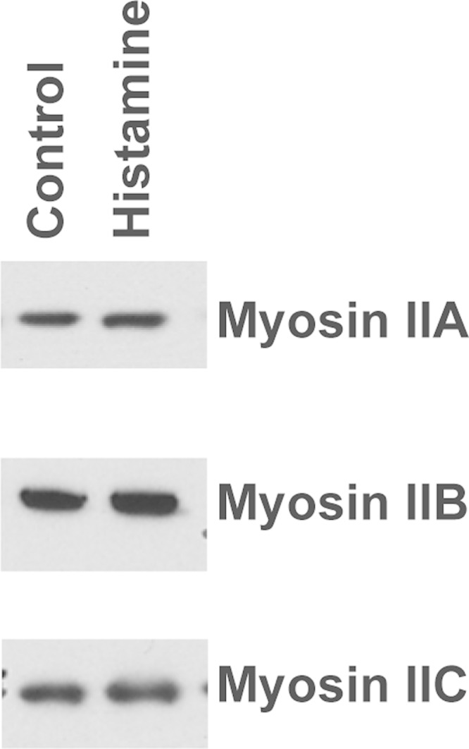
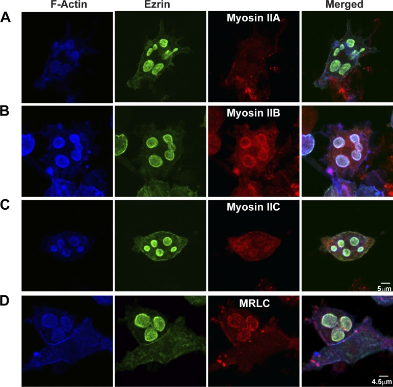
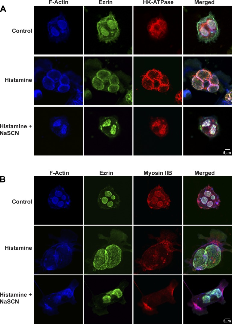
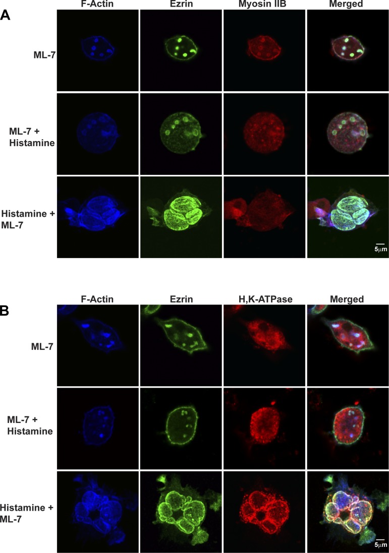
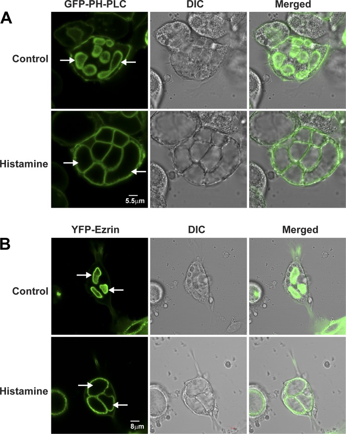
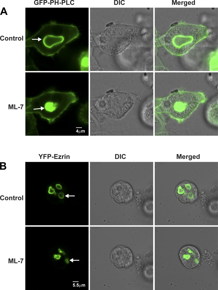
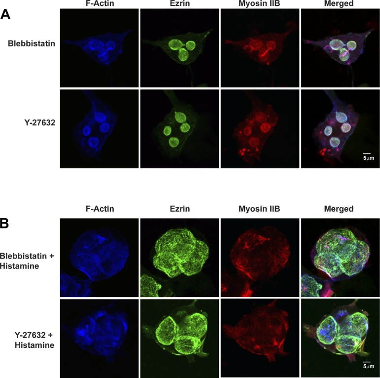
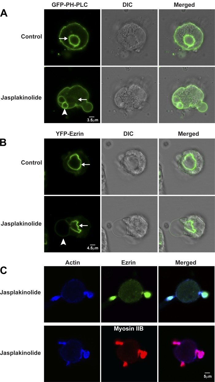
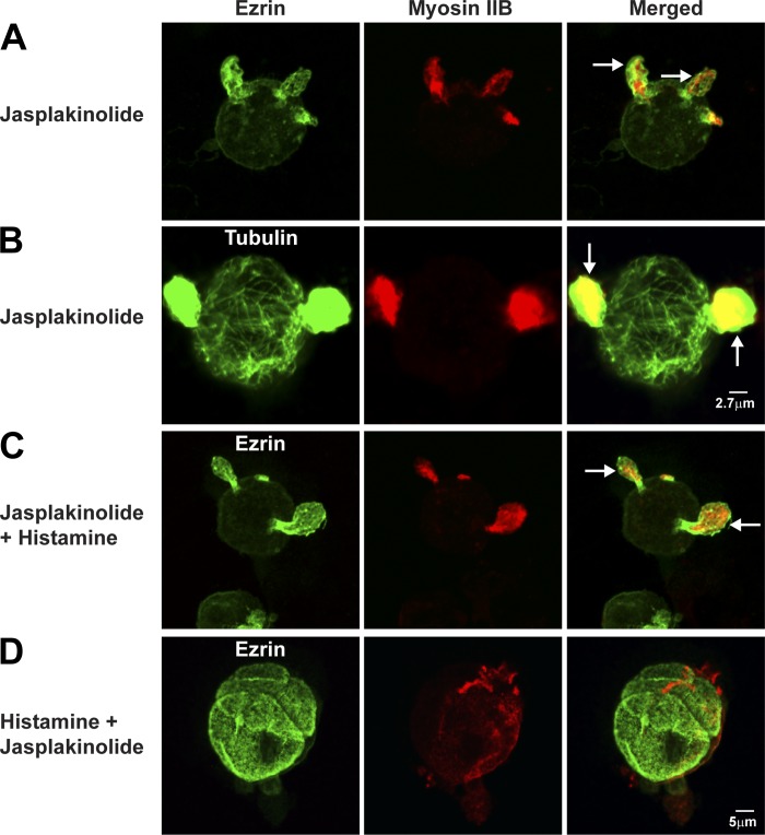
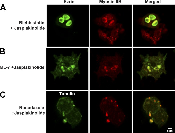
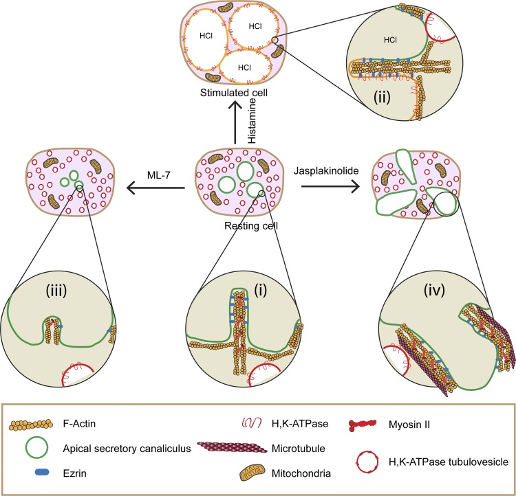
Selective inhibitors of myosin or actin function and confocal microscopy were used to test the role of an actomyosin complex in controlling morphology, trafficking, and fusion of tubulovesicles (TV) containing H-K-ATPase with the apical secretory canaliculus (ASC) of primary-cultured rabbit gastric parietal cells. In resting cells, myosin IIB and IIC, ezrin, and F-actin were associated with ASC, whereas H-K-ATPase localized to intracellular TV. Histamine caused fusion of TV with ASC and subsequent expansion resulting from HCl and water secretion; F-actin and ezrin remained associated with ASC whereas myosin IIB and IIC appeared to dissociate from ASC and relocalize to the cytoplasm. ML-7 (inhibits myosin light chain kinase) caused ASC of resting cells to collapse and most myosin IIB, F-actin, and ezrin to dissociate from ASC. TV were unaffected by ML-7. Jasplakinolide (stabilizes F-actin) caused ASC to develop large blebs to which actin, myosin II, and ezrin, as well as tubulin, were prominently localized. When added prior to stimulation, ML-7 and jasplakinolide prevented normal histamine-stimulated transformations of ASC/TV and the cytoskeleton, but they did not affect cells that had been previously stimulated with histamine. These results indicate that dynamic pools of actomyosin are required for maintenance of ASC structure in resting cells and for trafficking of TV to ASC during histamine stimulation. However, the dynamic pools of actomyosin are not required once the histamine-stimulated transformation of TV/ASC and cytoskeleton has occurred. These results also show that vesicle trafficking in parietal cells shares mechanisms with similar processes in renal collecting duct cells, neuronal synapses, and skeletal muscle.

Keywords: F-actin; membrane dynamics; nonmuscle myosin II; vesicle trafficking.


Figures

Fig. 1.
Fig. 1.
Expression of myosin II isoforms. Western blot of myosin IIA, IIB, and IIC in parietal cells. Isolated cells from rabbit stomachs were treated with either 100 μM cimetidine (Control) or 100 μM histamine (Histamine) for 30 min, then processed for Western blots to assay myosin heavy chain. Equal amounts of protein were loaded onto 4–12% SDS-PAGE gels. Proteins were transferred onto membranes and probed with antibodies to myosin heavy chain A, B, or C. Results were visualized with chemiluminescence reagent. There were no differences in myosin band intensities between control and histamine-stimulated cells.
Fig. 2.
Fig. 2.
Localization of myosin II, MRLC, actin, and ezrin in resting parietal cells. F-actin was stained by phalloidin conjugated with Alexa Fluor 647. Immunostaining was performed on ezrin with mouse anti-ezrin and myosin II by use of either rabbit anti-myosin IIA (A), rabbit anti-myosin IIB (B), rabbit anti-myosin IIC (C), or rabbit anti-MRLC (D) followed by Alexa Fluor-labeled goat secondary antibodies in resting parietal cells. Myosin IIB and IIC and MRLC colocalized with actin and ezrin in apical secretory canaliculus (ASC), but myosin IIA was localized near the basolateral membrane in resting parietal cells.
Fig. 3.
Fig. 3.
Effects of histamine and histamine + NaSCN on localization of myosin IIB, actin, ezrin, and H-K-ATPase in parietal cells. Parietal cells were treated with cimetidine (100 μM) (Control), histamine (100 μM), or histamine + NaSCN (3 mM NaSCN) together for 30 min, followed by fixation and staining for F-actin and ezrin as described in Fig. 2 and H-K-ATPase with mouse anti-H-K-ATPase (2G11) followed by Alexa Fluor-labeled goat secondary antibody (A) or F-actin, ezrin, and myosin IIB (B). F-actin and ezrin by were localized to ASCs in both resting and histamine-stimulated cells. Myosin IIB was localized to ASC in resting parietal cells and to the cytosol in histamine-stimulated cells. H-K-ATPase localized to tubulovesicles (TV) in cytosol in resting cells and to expanded ASC in histamine-stimulated cells. Cells treated with histamine + NaSCN together showed collapsed ASC, and ezrin, actin, and H-K-ATPase remained associated with the shrunken ASC, and myosin IIB remained dispersed to the cytosol. The less distinct staining of ezrin in images also stained for H-K-ATPase resulted from nonspecific binding of polyclonal ezrin antibody (i.e., compared with monoclonal antibody used in other images).
Fig. 4.
Fig. 4.
Effects of ML-7 on actin, ezrin, myosin IIB, and ASC morphology in resting and histamine-stimulated parietal cells. Cells were treated with cimetidine (100 μM) followed by ML-7 (10 μM) for 30 min or ML-7 for 30 min followed by histamine (100 μM) for 30 min (ML-7 + Histamine) or with histamine for 30 min followed by ML-7 for 30 min (Histamine + ML-7). Cells were then fixed and stained for F-actin, ezrin, and myosin IIB (A) or F-actin, ezrin, and H-K-ATPase (B) as described in legends to Figs. 2 and 3.
Fig. 5.
Fig. 5.
Localization of green fluorescent protein (GFP)-PLC1δ-PH and ezrin tagged with yellow fluorescent protein (YFP-ezrin) in live parietal cells: control and histamine stimulation. Primary cultures of parietal cells were infected with adenovirus (rAD) expressing GFP-PLC1δ-PH (A) and YFP-ezrin (B) 2 h after plating. Cells were maintained for 40 h at 37°C and treated for 30 min with either cimetidine (100 μM) to assure the resting state (Control) or with histamine (100 μM) to induce HCl secretion (Histamine). Fluorescence and differential interference (DIC) images are shown for the same cells in both control and histamine-treated states. Apical membrane vacuoles (arrows) became enlarged and occupied most of the cytoplasm in the stimulated compared with the resting state. GFP-PLC1δ-PH was localized to both ASC and basolateral membrane, whereas YFP-ezrin localized primarily to ASC membrane in resting and stimulated cells.
Fig. 6.
Fig. 6.
ML-7 causes ASC to shrink in resting parietal cells. Live cell fluorescence and DIC imaging of parietal cells expressing GFP-PLC1δ-PH (A) and YFP-ezrin (B) during treatment with 0.1% DMSO (Control) or ML-7 (10 μM) for 30 min. GFP-PLC1δ-PH and ezrin both localized to ASCs (arrows), which became shrunken/shriveled in the ML-7-treated compared with control cells.
Fig. 7.
Fig. 7.
Blebbistatin and Y-27632 do not affect ASC morphology of resting parietal cells or responses of parietal cells to histamine. A: resting parietal cells were treated with blebbistatin (50 μM) or Y-27632 (10 μM) for 30 min followed by immunostaining for F-actin, ezrin, and myosin IIB as described in Fig. 2. B: parietal cells were treated with blebbistatin or Y-27632 for 30 min, then stimulated with histamine (100 μM) for 30 min followed by fixation and immunostaining for F-actin, ezrin, and myosin IIB as described in Fig. 2.
Fig. 8.
Fig. 8.
Jasplakinolide causes blebbing and shrinkage of ASC membranes: localization of GFP-PLC1δ-PH and YFP-ezrin in control and jasplakinolide-treated live parietal cells. Parietal cells expressing GFP-PLC1δ-PH (A) and YFP-ezrin (B) were treated with 0.3% DMSO (Control) or jasplakinolide (3 μM) for 30 min. Fluorescence and corresponding DIC images are shown. Jasplakinolide caused membrane blebbing (arrowhead) from 1 part of ASC, which appeared to shrink (arrow) compared with the resting state. C: parietal cells were incubated with 3 μM jasplakinolide for 30 min followed by fixation and staining for actin using mouse anti-actin antibody and Alexa Fluor-labeled goat secondary antibody; ezrin and myosin IIB were labeled as described in Fig. 3. Actin colocalized with ezrin (top) and myosin IIB (bottom).
Fig. 9.
Fig. 9.
Jasplakinolide alters the localization of ezrin and myosin and morphology of ASC and inhibits histamine-stimulated ASC membrane expansion. Parietal cells were treated with jasplakinolide (3 μM) for 60 min (A and B) or jasplakinolide for 30 min followed by histamine (100 μM) for 30 min (Jasplakinolide + Histamine) (C) or with histamine for 30 min followed by jasplakinolide for 30 min (Histamine + Jasplakinolide) (D); cells were then fixed and stained for ezrin and myosin IIB as described in Fig. 2 (A, C, and D) or tubulin by using mouse anti-tubulin followed by Alexa Fluor-labeled goat secondary antibody (B). Arrows indicate membrane protrusion containing ezrin, myosin IIB, and tubulin.
Fig. 10.
Fig. 10.
Effects of combinations of jasplakinolide, blebbistatin, ML-7, and nocodazole on parietal cell ezrin, myosin IIB, and tubulin. Parietal cells were treated with blebbistatin (50 μM) for 30 min followed by jasplakinolide (3 μM) for another 30 min (Blebbistatin + Jasplakinolide) (A) or with ML-7 (10 μM) for 30 min followed by jasplakinolide for 30 min (ML-7 + Jasplakinolide) (B) or with nocodazole (20 μM) for 4 h followed by jasplakinolide for 30 min (Nocodazole + Jasplakinolide) (C); cells were then fixed and stained for ezrin, myosin IIB, and tubulin as described in Fig. 9.
Fig. 11.
Fig. 11.
Model for role of actomyosin complex in ASC morphology and TV trafficking in parietal cells. i: F-actin and myosin IIB assembled into filaments form an actomyosin complex that surrounds ASC in resting parietal cells; F-actin is attached to the ASC (green membrane) by ezrin. H-K-ATPase-containing TV (red membranes) are localized to the cytoplasm. ii: Histamine stimulation causes myosin IIB to dissociate from the ASC, whereas TV translocate from the cytosol to and fuse with ASC (which become yellow to denote fusion and mixing of TV with ASC). This fusion event is associated with activation of H-K-ATPase (and associated ion channels, not shown), leading to secretion of HCl and enlargement of ASC. F-actin and ezrin remain associated with ASC whereas myosin IIB dissociates from ASC in histamine-stimulated cells. iii: ML-7 causes ASC to shrink/collapse owing to F-actin depolymerization and dissociation of ezrin and myosin from ASC; these effects also prevent normal activation by histamine. iv: Jasplakinolide leads to stabilization of F-actin, which lengthens and, in association with myosin and microtubules, helps to form bleblike membrane protrusions of ASC that then prevent normal trafficking and fusion of TV with ASC. This effect of jasplakinolide is blocked when cells are pretreated with ML-7 or blebbistatin or nocodazole, indicating a role for myosin IIB and microtubules in the formation of the blebs.

Similar articles

Cited by

References

    1. Agnew BJ, Duman JG, Watson CL, Coling DE, Forte JG. Cytological transformations associated with parietal cell stimulation: critical steps in the activation cascade. J Cell Sci 112: 2639–2646, 1999. - PubMed
    1. Akagi K, Nagao T, Urushidani T. Responsiveness of beta-escin-permeabilized rabbit gastric gland model: effects of functional peptide fragments. Am J Physiol Gastrointest Liver Physiol 277: G736–G744, 1999. - PubMed
    1. Algrain M, Turunen O, Vaheri A, Louvard D, Arpin M. Ezrin contains cytoskeleton and membrane binding domains accounting for its proposed role as a membrane-cytoskeletal linker. J Cell Biol 120: 129–139, 1993. - PMC - PubMed
    1. Ammar DA, Nguyen PN, Forte JG. Functionally distinct pools of actin in secretory cells. Am J Physiol Cell Physiol 281: C407–C417, 2001. - PubMed
    1. Andzelm MM, Chen X, Krzewski K, Orange JS, Strominger JL. Myosin IIA is required for cytolytic granule exocytosis in human NK cells. J Exp Med 204: 2285–2291, 2007. - PMC - PubMed

Publication types

MeSH terms

LinkOut - more resources