. 2014 Jun;34(6):1249-59.
doi: 10.1161/ATVBAHA.114.303240. Epub 2014 Feb 27.

Identification and initial functional characterization of a human vascular cell-enriched long noncoding RNA

Affiliations

Identification and initial functional characterization of a human vascular cell-enriched long noncoding RNA

Robert D Bell et al. Arterioscler Thromb Vasc Biol. 2014 Jun.

Abstract

Objective: Long noncoding RNAs (lncRNAs) represent a rapidly growing class of RNA genes with functions related primarily to transcriptional and post-transcriptional control of gene expression. There is a paucity of information about lncRNA expression and function in human vascular cells. Thus, we set out to identify novel lncRNA genes in human vascular smooth muscle cells and to gain insight into their role in the control of smooth muscle cell phenotypes.

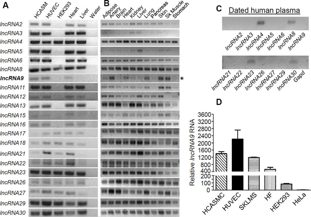
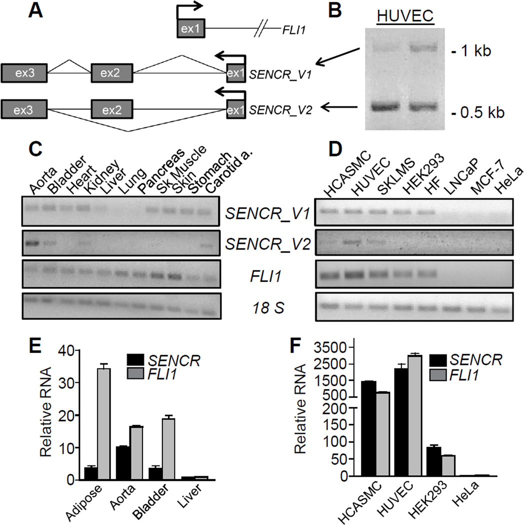
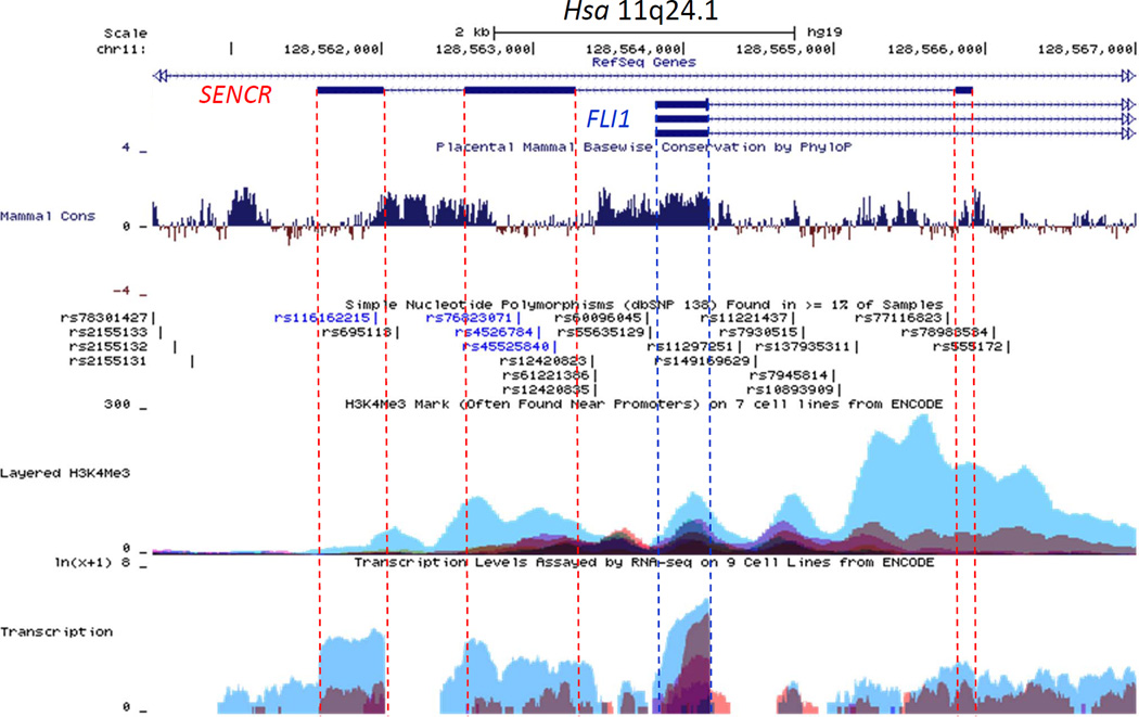
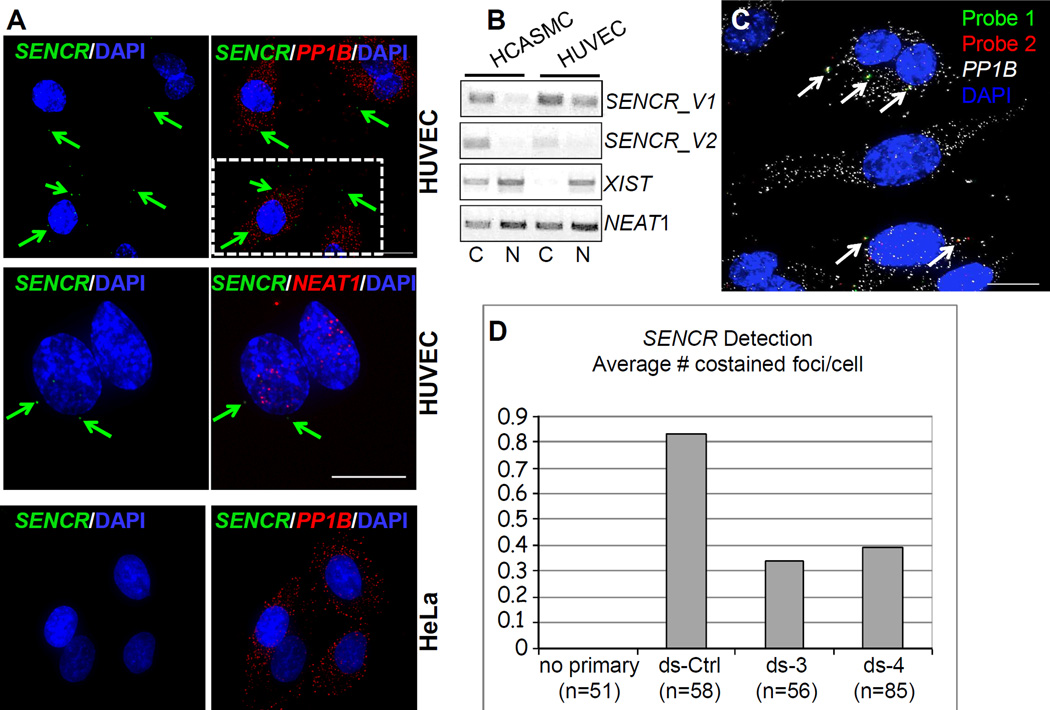
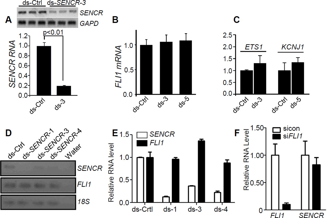
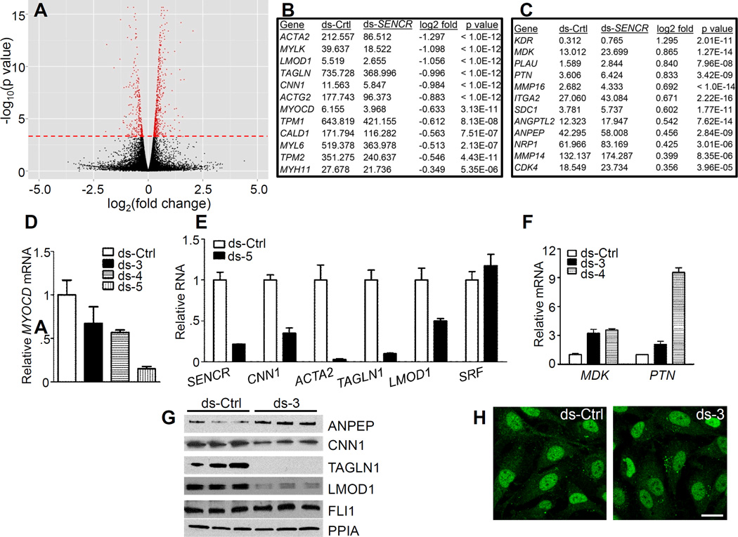
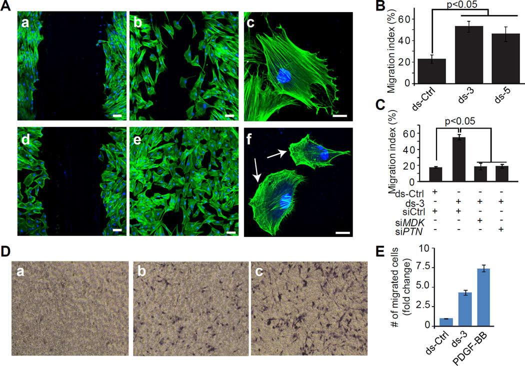
Approach and results: RNA sequencing (RNA-seq) of human coronary artery smooth muscle cells revealed 31 unannotated lncRNAs, including a vascular cell-enriched lncRNA (Smooth muscle and Endothelial cell-enriched migration/differentiation-associated long NonCoding RNA [SENCR]). Strand-specific reverse transcription polymerase chain reaction (PCR) and rapid amplification of cDNA ends indicate that SENCR is transcribed antisense from the 5' end of the FLI1 gene and exists as 2 splice variants. RNA fluorescence in situ hybridization and biochemical fractionation studies demonstrate SENCR is a cytoplasmic lncRNA. Consistent with this observation, knockdown studies reveal little to no cis-acting effect of SENCR on FLI1 or neighboring gene expression. RNA-seq experiments in smooth muscle cells after SENCR knockdown disclose decreased expression of Myocardin and numerous smooth muscle contractile genes, whereas several promigratory genes are increased. Reverse transcription PCR and Western blotting experiments validate several differentially expressed genes after SENCR knockdown. Loss-of-function studies in scratch wound and Boyden chamber assays support SENCR as an inhibitor of smooth muscle cell migration.

Conclusions: SENCR is a new vascular cell-enriched, cytoplasmic lncRNA that seems to stabilize the smooth muscle cell contractile phenotype.

Keywords: RNA sequence; RNA, long noncoding; cell migration; endothelial cells; myocytes, smooth muscle.


Figures

Figure 1
Figure 1. Validation of lncRNA expression in human cells and tissues
RT-PCR analysis of 21 lncRNAs (arbitrarily numbered) in indicated human cells (A) and tissues (B). Bold lncRNA9 and asterisk denote SENCR. (C) RT-PCR of indicated lncRNAs in dated human plasma. All reactions were done using the same PCR parameters. (D) Quantitative RT-PCR of lncRNA9 in the indicated human cell types.
Figure 2
Figure 2. SENCR gene structure and isoform expression
(A) Schematic of SENCR and FLI1 (partial) gene loci. Arrows denote the transcription start sites and bent lines in SENCR indicate splicing patterns. (B) RT-PCR of SENCR with primers to exon 1 and 3 showing the presence of two transcripts reflecting full length (V1) and alternately spliced (V2) SENCR. RT-PCR of two SENCR isoforms and FLI1 in various human tissues (C) and cell lines (D). Quantitative RT-PCR of SENCR and FLI1 in select human tissues (E) and cell lines (F). Bars here and below represent the standard deviation of one experiment with three biological replicates. All expression data here and below represent at least two (more typically multiple) independent studies performed by more than one author. Unless indicated otherwise, SENCR expression here and below reflects both isoforms using primers to a common exon. SKLMS, uterine leiomyosarcoma cell line; HF, human fibroblasts.
Figure 3
Figure 3. UCSC genome browser track of the human FLI1-SENCR sense-antisense gene pair
The SENCR gene comprises 3 exons (shown as dark rectangles) and 2 introns. The first exon of SENCR initiates on the opposite strand ~1.5 kb downstream from the first exon of FLI1. The dotted vertical lines serve to highlight several features, including (from the top) mammalian conservation (Mammal Cons), reference SNPs (rs numbers), H3K4Me3, and RNA-seq (Transcription) in Tier 1 and Tier 2 cells from ENCODE. Note the lower conservation and selective transcription in HUVEC (blue peaks at bottom) for SENCR as compared to FLI1. See Results for more details.
Figure 4
Figure 4. Localization of SENCR RNA
(A) RNA FISH analysis of SENCR versus cytoplasmic PP1B mRNA and nuclear NEAT1 RNA in indicated cell types. Arrows point to single molecules of SENCR RNA in the cytoplasm of HUVEC. Scale bars are all 5 µm. The broken white rectangle at upper right is shown at higher magnification in Figure III in the online only Data supplement. (B) RT-PCR analysis of two SENCR isoforms versus other lncRNA genes from polyA+ RNA isolated from the cytoplasmic (C) or nuclear (N) fractions of indicated cell types. (C) Application of two probe sets to SENCR RNA (see Methods). Arrows point to coincident localization of two fluorescently tagged probe sets (yellow) targeting different regions of the SENCR transcript. (D) Quantitative measures of coincident localization of SENCR probes in HUVEC transfected with a dicer substrate control RNA (ds-Ctrl) or two dicer substrate RNAs targeting different regions of SENCR (ds-3 and ds-4). The Y-axis indicates the average number of co-stained foci/cell.
Figure 5
Figure 5. Effect of knocking down SENCR on local gene expression
(A) Dicer substrate control (ds-Ctrl) or dicer substrate SENCR RNA was transfected into HCASMC for 72 hr and then total RNA isolated for conventional (top) or quantitative (bottom) RT-PCR. Dicer substrate RNA to various regions of SENCR are abbreviated here and below as “ds” followed by a number (see Table I in the online only Data supplement for details). Quantitative RT-PCR of FLI1 mRNA (B) or flanking genes around FLI1 (C) following 3 d transfection with indicated dicer substrate RNAs. (D) Conventional RT-PCR of SENCR and FLI1 in HUVEC following transfection with indicated dsRNAs. (E) Quantitative RT-PCR of SENCR and FLI1 following transfection with indicated dsRNAs in HUVEC. (F) Quantitative RT-PCR of FLI1 and SENCR following knockdown of FLI1 mRNA in HCASMC. Data are representative of multiple independent experiments carried out by independent investigators using several isolates of HCASMC or HUVEC.
Figure 6
Figure 6. Effect of SENCR knockdown on HCASMC transcriptome
(A) Volcano plot depicting changes in gene expression with SENCR knockdown. The red dashed line indicates genes (in red) whose changes in expression were statistically significant. Sample SMC contractile genes (B) and pro-migratory genes (C) exhibiting either reduced (B) or increased (C) expression with ds-SENCR knockdown. See Table 3 in the online only Data supplement for a complete listing of all genes showing significant up- or down-regulation with SENCR knockdown. Quantitative RT-PCR validation of reduced MYOCD mRNA (D) and SMC contractile genes (E) in HCASMC following knockdown of SENCR with various dsRNAs. (F) Quantitative RT-PCR validation of up-regulation of two pro-migratory genes upon knockdown of SENCR in HCASMC. (G) Western blot validation of up-regulated (ANPEP) and down-regulated (SMC contractile) proteins in HCASMC 72 hr after indicated transfection with dsRNA. Similar findings were observed in an independent experiment. (H) Immunofluorescence microscopy of FLI1 protein in the nucleus of HCASMC following 3 d transfection with indicated dsRNAs. Results are representative of multiple experiments performed by independent investigators. Scale bar = 10 µm for both images.
Figure 7
Figure 7. Effect of SENCR knockdown in a scratch wound assay of cell migration
(A) HCASMC were transfected with ds-Ctrl (panels a–c) or ds-SENCR-3 (panels d-f) for 72 hr after which a scratch wound was created and cell migration assessed in quiescent (panels a, d) HCASMC or in similar cells stimulated for 12 hr with 10% FBS (panels b, c, e, f). Cells were stained with phalloidin (red) and DAPI (blue). Bars are 25 µm in panels a, b, d, and e and 10 µm in panels c and f. Arrows in panel f denote lamellipodia. (B) Quantitative measure of the area of the wound occupied by dsRNA-transfected HCASMC 12 hrs following serum stimulation. (C) Same experiment as in B only HCASMC were transfected simultaneously with a control siRNA (siCtrl) or an siRNA to one of two pro-migratory genes. Each siRNA reduced level of MDK or PTN mRNA by more than 80% (Figure V in the online only Data supplement). (D) Effect of SENCR knockdown on HCASMC migration in a Boyden chamber. Cells were transfected with ds-Ctrl (a), ds-3 (b) or 25 ng/ml PDGF-BB (c) for 6 hr and the fold change in number of cells migrating through the porous membrane quantitated (E). The data reflect cell counts from 5 independent fields.

Comment in

  • The smooth long noncoding RNA SENCR.
    Thum T, Kumarswamy R. Thum T, et al. Arterioscler Thromb Vasc Biol. 2014 Jun;34(6):1124-5. doi: 10.1161/ATVBAHA.114.303504. Arterioscler Thromb Vasc Biol. 2014. PMID: 24828518 No abstract available.

Similar articles

Cited by

References

    1. Ohno S. So much "junk" DNA in our genome. Brookhaven Symp Biol. 1972;23:366–370. - PubMed
    1. Okazaki Y, Furuno M, Kasukawa T, et al. Analysis of the mouse transcriptome based on functional annotation of 60,770 full-length cdnas. Nature. 2002;420:563–573. - PubMed
    1. Kapranov P, Cawley SE, Drenkow J, Bekiranov S, Strausberg RL, Fodor SPA, Gingeras TR. Large-scale transcriptional activity in chromosomes 21 and 22. Science. 2002;296:916–919. - PubMed
    1. Carninci P, Kasukawa T, Katayama S, et al. The transcriptional landscape of the mammalian genome. Science. 2005;309:1559–1563. - PubMed
    1. Cheng J, Kapranov P, Drenkow J, et al. Transcriptional maps of 10 human chromosomes at 5-nucleotide resolution. Science. 2005;308:1149–1154. - PubMed

Publication types

MeSH terms