. 2014 Jun;13(6):1397-411.
doi: 10.1074/mcp.M113.034108. Epub 2014 Feb 27.

The Vac14-interaction network is linked to regulators of the endolysosomal and autophagic pathway

Affiliations

The Vac14-interaction network is linked to regulators of the endolysosomal and autophagic pathway

Ulf Schulze et al. Mol Cell Proteomics. 2014 Jun.

Abstract

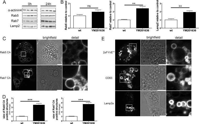
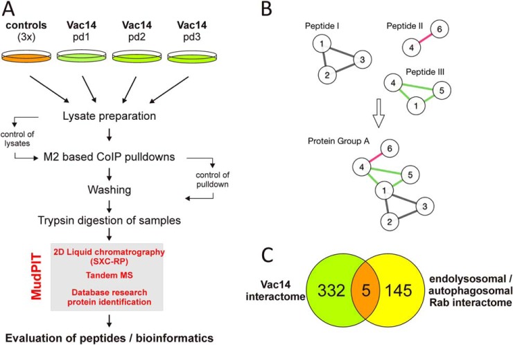
The scaffold protein Vac14 acts in a complex with the lipid kinase PIKfyve and its counteracting phosphatase FIG4, regulating the interconversion of phosphatidylinositol-3-phosphate to phosphatidylinositol-3,5-bisphosphate. Dysfunctional Vac14 mutants, a deficiency of one of the Vac14 complex components, or inhibition of PIKfyve enzymatic activity results in the formation of large vacuoles in cells. How these vacuoles are generated and which processes are involved are only poorly understood. Here we show that ectopic overexpression of wild-type Vac14 as well as of the PIKfyve-binding deficient Vac14 L156R mutant causes vacuoles. Vac14-dependent vacuoles and PIKfyve inhibitor-dependent vacuoles resulted in elevated levels of late endosomal, lysosomal, and autophagy-associated proteins. However, only late endosomal marker proteins were bound to the membranes of these enlarged vacuoles. In order to decipher the linkage between the Vac14 complex and regulators of the endolysosomal pathway, a protein affinity approach combined with multidimensional protein identification technology was conducted, and novel molecular links were unraveled. We found and verified the interaction of Rab9 and the Rab7 GAP TBC1D15 with Vac14. The identified Rab-related interaction partners support the theory that the regulation of vesicular transport processes and phosphatidylinositol-modifying enzymes are tightly interconnected.


Figures

Fig. 1.
Fig. 1.
Vac14 overexpression causes vacuolization. With the use of CellTracker blue in live cell imaging experiments, vacuoles were clearly distinguished from the blue labeled cytoplasm. A, both overexpression of a 3xFLAG Vac14 wt and the PIKfyve-binding deficient 3xFLAG Vac14 L156R mutant result in strong vacuolization of HeLa cells. Vacuoles are almost absent in the control cells (Ctrl) expressing the 3xFLAG tag alone. B, vacuoles caused by PIKfyve inhibitor YM201636 (24 h, 800 nm) versus untreated cells (Ctrl). C–E, quantification of the vacuoles illustrated in A and B, concerning (C) the amount of vacuoles per cell, (D) the vacuolar area as a percentage per cell, and (E) the size distribution of vacuoles caused by YM201636 treatment or Vac14 wt and Vac14 L156R overexpression, respectively. Detail shows the magnification of the indexed areas. Bar: 20 μm; differential interference contrast (DIC).
Fig. 2.
Fig. 2.
Vac14-associated vacuoles accumulate endolysosomal markers. Lysates of 3xFLAG Vac14 overexpressing HeLa cells (wt and L156R) were analyzed via quantitative Western blotting and compared with control lysates. Both Vac14 overexpressing approaches (wt and L156R) resulted in a significant increase of endogenous Rab7-, CD63-, and Lamp2-positive structures. The increase was stronger in Vac14 L156R lysates. A, sets of three independent quantifications of the Western blots were normalized to the loading control GAPDH and α-actinin 4. In all experiments, cell lines with an empty 3xFLAG plasmid served as a control. B, quantitative evaluation of at least eight samples from the proteins shown in A. ns = not significant; *p < 0.05, **p < 0.01, ***p < 0.001.
Fig. 3.
Fig. 3.
Vac14 overexpression enlarges compartments of the endolysosomal pathway. Vac14-overexpressing cells were transiently transfected with fluorescent-tagged marker proteins in order to examine the vacuole in more detail. Stable cell lines with an empty 3xFLAG plasmid (A) or expressing 3xFLAG-tagged Vac14 wt (B) and L156R mutant (C) were transiently transfected with plasmids encoding constitutively active EGFP-Rab5 and -Rab7, EGFP-2xFYVEhrs, EGFP-CD63, and Lamp2-mCherry. Vac14-associated vacuoles were predominantly positive for late endosomal marker proteins (Rab7, CD63, or Lamp2). D, quantitative analysis of EGFP/mCherry-positive compartments. The size of EGFP/mCherry protein structures (panels A–C) in Vac14-overexpressing cells (wt and L156R) is shown as the fold increase relative to the control (Ctrl; empty 3xFLAG plasmid). ns = not significant; *p < 0.05, **p < 0.01, ***p < 0.001. Size bar and detailed view: 10 μm.
Fig. 4.
Fig. 4.
Inhibition of PIKfyve kinase and Vac14 overexpression cause similar effects. A, HeLa cells incubated with PIKfyve inhibitor YM201636 (800 nm; 24 h) showed a significant accumulation of endogenous Rab7 and Lamp2 but not of Rab5. α-actinin 4 served as a loading control. Sets of four protein samples from control cells (left) or treated cells (right) are shown. B, quantitative densitometric analysis of Western blots shown in A; quantifications of the Western blots were normalized to the loading control α-actinin 4. C, live cell imaging analysis of HeLa cells treated with YM201636 showed that the vacuoles were positive for Rab5 and Rab7. D, quantification of Rab5- and Rab7-positive structures showed significant enlargement (up to 3.5-fold) of Rab5 and Rab7 structures in YM201636-stimulated cells. E, the limiting membrane of these vacuoles was also positive for CD63, Lamp2, and PI3P lipids (EGFP 2xFYVEhrs). ns = not significant; **p < 0.01, ***p < 0.001. Size bar and detailed view: 10 μm.
Fig. 5.
Fig. 5.
Vac14-complex-associated vacuolization causes an accumulation of autophagy marker proteins. Vacuolization due to Vac14 overexpression or YM201636 treatment was accompanied by a significant accumulation of autophagy marker proteins p62 and LC3-II. A, overexpression of Vac14 (wt and L156R) showed an increased level of p62 and LC3-II relative to the control cells (three samples each set). Densitometric evaluation of the Western blot analysis is shown on the right. B, lysates from HeLa cells treated with the PIKfyve inhibitor YM201636 for 24 h compared with nontreated cells. YM201636-derived vacuolization resulted in an accumulation of p62 and LC3-II. GAPDH served as a loading control. *p < 0.05, **p < 0.01, ***p < 0.001.
Fig. 6.
Fig. 6.
Schematic overview of the used MudPIT strategy. A, an overview of the used strategy is shown in the workflow scheme. Three independent pulldown assays in HEK293 cells expressing 3xFLAG Vac14 wt or 3xFLAG alone were used for the MudPIT assays. The CoIP/pulldown assays (pd1–pd3 and controls) were performed with beads covalently linked to anti-FLAG M2 antibodies. To verify successful coupling of FLAG-Vac14 to the M2-beads, aliquots of the CoIP/pulldown samples were controlled by SDS-PAGE and Western blot or Coomassie Blue staining (not shown). After stringency washings, protein samples were digested with trypsin and analyzed via the MudPIT technique, including two-dimensional LC and tandem MS (box). B, illustration of an evaluation example of protein groups and their localization tags. Proteins are shown as nodes, and peptides are shown as edges. Peptide I, shared by proteins 1, 2, and 3, was found in the pulldown (PD) and the control (C), and therefore the edges are colored gray. Peptide II, shared by proteins 4 and 6, was found in C only (red). Peptide III was found in PD only and was shared by proteins 1, 4, and 5 (green). Iterative merging of all protein groups sharing proteins resulted in protein group A, which showed a mixed localization tag, and thus none of those proteins could be incorporated in further analysis. However, if all edges/peptides were green in the final group, then those proteins could be incorporated into further analysis (supplemental information SI 1 and SI 2). C, analysis of the Vac14 interactome resulted in 337 proteins reviewed by the UniProtKB/Swiss-Prot databases. These were compared with the Rab family members and its interaction partners displayed in supplemental Table S1. TBC1D15, Rab9, SUN2, IDH3A, and NSF represent the intersection of both networks.
Fig. 7.
Fig. 7.
Vac14 interacts with the Rab7 GAP TBC1D15 and Rab9. A, stable 3xFLAG-overexpressing cell lines were transiently transfected with myc-TBC1D15. Beads that were covalently linked to the FLAG-tag specific antibody M2 were used in CoIP assays. Only lysates that expressed 3xFLAG-Vac14 wt or the Vac14 L156R mutant showed a direct association with co-expressed myc-TBC1D15 and endogenous Rab9. Cells expressing myc-TBC1D15 and the 3xFLAG alone served as negative controls and were not able to precipitate myc-TBC1D15 and endogenous Rab9. B, overexpression of 3xFLAG-Vac14 wt and Vac14 L156R in HeLa cells led to a significant accumulation of Rab9 relative to the loading control GAPDH. C, the same effect was observed in cells treated with the PIKfyve inhibitor YM201636 when compared with the loading control GAPDH (Fig. 5B). D, EGFP-Rab9 CA localized on the limiting membranes of vacuoles induced by overexpression of Vac14 wt/L156R or YM201636 treatment. *p < 0.05, ***p < 0.001. Size bar and detailed view: 10 μm.

References

    1. Di P. G., De C. P. (2006) Phosphoinositides in cell regulation and membrane dynamics. Nature 443, 651–657 - PubMed
    1. Mayinger P. (2012) Phosphoinositides and vesicular membrane traffic. Biochim. Biophys. Acta 1821, 1104–1113 - PMC - PubMed
    1. McCartney A. J., Zhang Y., Weisman L. S. (2014) Phosphatidylinositol 3,5-bisphosphate: low abundance, high significance. Bioessays 36, 52–64 - PMC - PubMed
    1. Shisheva A. (2012) PIKfyve and its lipid products in health and in sickness. Curr. Top. Microbiol. Immunol. 362, 127–162 - PubMed
    1. Jin N., Chow C. Y., Liu L., Zolov S. N., Bronson R., Davisson M., Petersen J. L., Zhang Y., Park S., Duex J. E., Goldowitz D., Meisler M. H., Weisman L. S. (2008) VAC14 nucleates a protein complex essential for the acute interconversion of PI3P and PI(3,5)P(2) in yeast and mouse. EMBO J. 27, 3221–3234 - PMC - PubMed

Publication types

MeSH terms

LinkOut - more resources