. 2014 Feb 28;343(6174):1002-5.
doi: 10.1126/science.1245831.

Promoter-bound trinucleotide repeat mRNA drives epigenetic silencing in fragile X syndrome

Affiliations

Promoter-bound trinucleotide repeat mRNA drives epigenetic silencing in fragile X syndrome

Dilek Colak et al. Science. .

Abstract

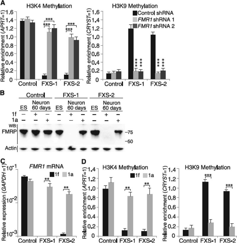
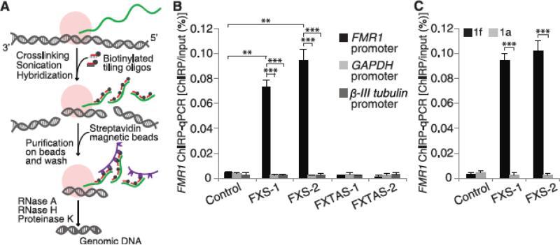
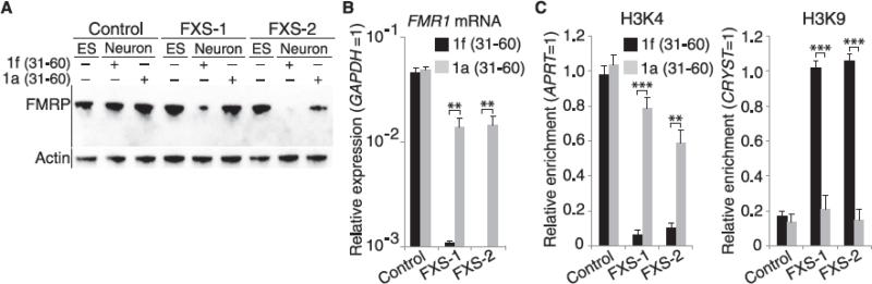
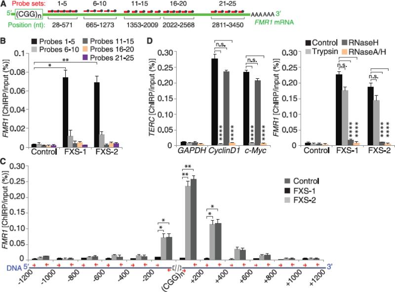
Epigenetic gene silencing is seen in several repeat-expansion diseases. In fragile X syndrome, the most common genetic form of mental retardation, a CGG trinucleotide-repeat expansion adjacent to the fragile X mental retardation 1 (FMR1) gene promoter results in its epigenetic silencing. Here, we show that FMR1 silencing is mediated by the FMR1 mRNA. The FMR1 mRNA contains the transcribed CGG-repeat tract as part of the 5' untranslated region, which hybridizes to the complementary CGG-repeat portion of the FMR1 gene to form an RNA·DNA duplex. Disrupting the interaction of the mRNA with the CGG-repeat portion of the FMR1 gene prevents promoter silencing. Thus, our data link trinucleotide-repeat expansion to a form of RNA-directed gene silencing mediated by direct interactions of the trinucleotide-repeat RNA and DNA.


Figures

Fig. 1
Fig. 1
The FMR1 transcript and its CGG-repeat tract are required for FMR1 silencing. (A) FMR1 mRNA is required for FMR1 silencing in differentiating FXS hESCs. shRNA-expressing lentivirus was applied at day 1, and histone marks at FMR1 promoters were measured at day 60. FXS hESCs expressing control shRNA showed high levels of transcriptionally repressive marks (H3K9me2) and low levels of transcriptionally active marks (H3K4me2). FMR1-specific shRNA prevented the appearance of repressive marks and maintained the expression of transcriptionally active marks (n = 4 per condition). ES, hESCs; WB, Western blot; APRT, adenine phosphoribosyltransferase; GAPDH, glyceraldehyde-3-phosphate dehydrogenase; CRYST, crystallin. (B to D) The CGG-repeat RNA-binding small molecule 1a blocks FMR1 silencing. Differentiating FXS hESCs treated with 10 μM 1a did not lose FMRP (B) or FMR1 mRNA (C) (n = 3 per condition) and retained active FMR1 promoters (D) (n = 3 per condition). Data are means ± SEM. Statistical analysis was performed using Student's t test (two-tailed distribution, **P < 0.01, ***P < 0.001). When comparing different cell lines, we considered the samples as two samples with unequal variance. When comparing different conditions on the same cell line, we considered the samples as two samples with equal variance.
Fig. 2
Fig. 2
FMR1 mRNA interacts with the FMR1 promoter in a CGG repeat–dependent manner. (A) Schematic representation of ChIRP technique [adapted from ()]. RNA (green) and protein complexes (pink) are cross-linked to the DNA (gray) in cells by glutaraldehyde. The cell lysate is sonicated to shear DNA to ~500 bp. Streptavidin beads (purple) are used to pull down biotinylated oligonucleotides hybridized to RNA. Bound DNA sequences are detected by qPCR. (B) FMR1 mRNA interacts with the FMR1 promoter. FMR1 mRNA bound to the FMR1 gene was measured by ChIRP at day 45 of differentiation (see fig. S11 for other time points). FMR1 mRNA was readily detectable on the FMR1 promoter in FXS neurons but not control neurons (n = 3 per condition). FMR1 mRNA does not bind to FMR1 promoters in FMR1 premutation lines, FXTAS-1 and FXTAS-2, that contain 70 and 73 CGG repeats, respectively. GAPDH and β-III tubulin promoters were used as controls. (C) The CGG-repeat portion of the transcript is required for the binding of FMR1 mRNA to the FMR1 gene. FMR1 binding to the FMR1 gene was markedly reduced in FXS hESC-derived neurons cultured in the presence of 1a. The control compound 1f did not block the FMR1 transcript–FMR1 gene interaction. Data are means ± SEM; Student's t test (two-tailed distribution, **P < 0.01, ***P < 0.001). Different conditions on the same cell line were considered as two samples with equal variance; different cell lines were considered as two samples with unequal variance.
Fig. 3
Fig. 3
Temporal requirement for FMR1 mRNA binding to the FMR1 promoter. (A) The small molecule 1a blocks the drop in FMRP levels during days 31 to 60 of differentiation. To determine when FMR1 mRNA is required for FMR1 silencing, we applied 1a to FXS hESCs during days 31 to 60 of differentiation. 1a maintained FMRP expression in FXS neurons. (B and C) 1a prevents FMR1 silencing. In FXS neurons, application of 1a during days 31 to 60 of differentiation was sufficient to maintain FMR1 mRNA levels [(B), quantitative reverse transcription polymerase chain reaction (qRT-PCR), n = 4 per condition], high levels of H3K4me2 and low levels of H3K9me2 histone modifications [(C), chromatin immunoprecipitation, n = 4 per condition]. Control hESCs were unaffected by 1a. Application of 1a (10 μM) during days 1 to 30 of differentiation failed to prevent FMR1 silencing (see fig. S12). Data are means ± SEM; Student's t test (two-tailed distribution, **P < 0.01, ***P < 0.001). Different conditions on the same cell line were considered as two samples with equal variance.
Fig. 4
Fig. 4
The CGG-repeat portion of the FMR1 mRNA hybridizes to the complementary region of the FMR1 DNA. (A) Schematic of the hybridization sites of the different biotinylated-oligonucleotide sets used to pull down sheared RNA in ChIRP experiments (see methods for further details on probe design). (B) The 5′ UTR portion of the FMR1 transcript binds to the FMR1 gene. ChIRP was performed using the probe sets shown in (A). Only probes that bind the CGG repeat–proximal portion of the FMR1 transcript pulled down the FMR1 promoter in FXS neurons (n = 4 per condition). (C) The FMR1 transcript binds to the CGG-repeat portion of the FMR1 gene. To determine where the FMR1 mRNA binds on the FMR1 gene, we measured the ChIRP signal along a 1200-bp region both upstream and downstream of the genomic CGG repeat. The positions of the primers used to amplify portions of the FMR1 gene (blue) are indicated (red arrows) (referred to as bp relative to the 5′ or 3′ end of the CGG repeat). The ChIRP signal was highly enriched adjacent to the genomic CGG repeat (n = 3 per condition) (see also fig. S16). (D) The FMR1 mRNA binds to the FMR1 DNA in a protein-independent and RNase H–sensitive manner. The binding of the control noncoding-RNA TERC to its target promoters (CyclinD1 and c-Myc) was abolished after trypsin treatment (n = 3), whereas the binding of the FMR1 transcript to the FMR1 gene was unaffected by trypsin in FXS hESC-derived neurons (n = 3 per condition). In contrast, RNase H treatment only blocked the FMR1 ChIRP signal (n = 3 per condition). RNase A–RNase H treatment, which digests all RNA, is used as a control to demonstrate the RNA-dependence of the ChIRP signal. Data are means ± SEM; Student's t test (two-tailed distribution, *P < 0.05, **P < 0.01, ****P < 0.0001). Different conditions on the same cell line were considered as two samples with equal variance; different cell lines were considered as two samples with unequal variance.

Similar articles

Cited by

References

    1. Verkerk AJ, et al. Cell. 1991;65:905–914. - PubMed
    1. Oberlé I, et al. Science. 1991;252:1097–1102. - PubMed
    1. Coffee B, Zhang F, Ceman S, Warren ST, Reines D. Am. J. Hum. Genet. 2002;71:923–932. - PMC - PubMed
    1. Willemsen R, Bontekoe CJ, Severijnen LA, Oostra BA. Hum. Genet. 2002;110:601–605. - PubMed
    1. Brouwer JR, et al. Exp. Cell Res. 2007;313:244–253. - PMC - PubMed

Publication types