. 2014 Feb 28;343(6174):1010-4.
doi: 10.1126/science.1249484.

Detection of a recurrent DNAJB1-PRKACA chimeric transcript in fibrolamellar hepatocellular carcinoma

Affiliations

Detection of a recurrent DNAJB1-PRKACA chimeric transcript in fibrolamellar hepatocellular carcinoma

Joshua N Honeyman et al. Science. .

Abstract

Fibrolamellar hepatocellular carcinoma (FL-HCC) is a rare liver tumor affecting adolescents and young adults with no history of primary liver disease or cirrhosis. We identified a chimeric transcript that is expressed in FL-HCC but not in adjacent normal liver and that arises as the result of a ~400-kilobase deletion on chromosome 19. The chimeric RNA is predicted to code for a protein containing the amino-terminal domain of DNAJB1, a homolog of the molecular chaperone DNAJ, fused in frame with PRKACA, the catalytic domain of protein kinase A. Immunoprecipitation and Western blot analyses confirmed that the chimeric protein is expressed in tumor tissue, and a cell culture assay indicated that it retains kinase activity. Evidence supporting the presence of the DNAJB1-PRKACA chimeric transcript in 100% of the FL-HCCs examined (15/15) suggests that this genetic alteration contributes to tumor pathogenesis.


Figures
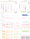
Fig. 1
Fig. 1. RNA-seq read coverage from fibrolamellar hepatocellular carcinoma and adjacent healthy liver tissue
(A-C) Plot of reads mapped to chromosome 19 in the region encoding on the negative strand the genes PRKACA (chr19:14,202,499 - 14,228,558) and DNAJB1 (chr19:14,625,580 - 14,640,086) from the normal tissue (blue) and FL-HCC tissue (red). (A) Normalized RNA-Seq read counts from nine pairs of tumor and adjacent tissue demonstrate a consistent increase in tumor relative to normal in the reads of exon 2–10 of PRKACA, and a decrease in the reads of exon 1. Normalized read counts are plotted per exon part (non-overlapping portions of exons in all isoforms in ENSEMBL annotation; indicated by empty grey boxes). Transcript structure (solid color boxes) indicates most likely dominant isoform as inferred by RNA-Seq read coverage. Lines indicate the average normalized read count per exon part (in dominant isoform) for normal and tumor samples. (B) Sashimi plot (39) of RNA-Seq read coverage at PRKACA and DNAJB1 loci for patient 9. Solid peaks depict reads per kilobase per million reads mapped (RPKM) within individual exons. Reads that bridge different exons are shown as arcs. In every tumor sample (9 out of 9) and in none of the normal tissue sample (0 out of 9), there are reads mapped from the end of exon 1 of DNAJB1 to the start of exon 2 of PRKACA. (C) There is an additional set of reads in patient 4 that map from the second exon of DNAJB1 to the start of the second exon of PRKACA. Indistinguishable results are observed in metastasis tissue from this same patient. (D) RNA-Seq read mapping predicts the production of four transcripts: a native DNAJB1 (green), a native isoform 1 PRKACA (orange), a predominant chimera with the first exon of DNAJB1 and exons 2–10 of PRKACA and, in a subset of patients, a minority transcript with the first exon and part of the second of DNAJB1 and exons 2–10 of PRKACA. (E). Sanger Sequencing of RT-PCR products from FL-HCC samples confirmed a chimera transcript in 7 out of 7 patients joining the end of exon 1 of DNAJB1 and the start of exon 2 of PRKACA.
Fig. 2
Fig. 2. DNA sequence analysis of fibrolamellar hepatocellular carcinoma DNA
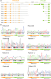
(A) Mapping of the size and location of the breaks in the DNA between the DNAJB1 and the PRKACA genes. (B). PCR followed by Sanger sequencing confirmed a deletion of ~400kD in each patient. Each deletion results in a fusion that starts either in intron 1 or exon 2 of DNAJB1 and ends in intron 1 of PRKACA. The break is in a different location in all patients. Note that the sequencing reads are shown off of the sense-strand. However, both DNAJB1 and PRKACA are coded off the negative strand.
Fig. 3
Fig. 3. Tumor-specific expression of a protein consistent with the DNAJB1-PRKACA chimera
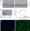
(A)Immunoblot analysis. Protein extracts of fibrolamellar carcinoma (T) and adjacent liver tissue (N) were separated on SDS-PAGE and subjected to immunoblot analysis using an antibody to the carboxyl-terminus of PRKACA. This analysis revealed the presence of the native PRKACA in all tumor, metastasis and normal samples and the presence of one additional, apparent higher molecular weight band in all tumor samples (the predominant chimera). There is a second even higher molecular weight band, the minority chimera, in the two tumor samples that had demonstrated a second set of RNA reads mapping between exon 2 of DNAJB1 and exon 2 of PRKACA (Patient 4, 14). (B) Confirmation of Chimeric Protein. Protein extracts of fibrolamellar carcinoma (T) and adjacent liver tissue (N) were immunoprecipitated with an antibody to the amino-terminus of DNAJB1 and run adjacent to total cell extract on SDS-PAGE. These samples were then subjected to immunoblot analysis using an antibody to the carboxyl-terminus of PRKACA (C) PKA activity of WT PRKACA and Chimera are indistinguishable. HEK-293T cells were transfected with an empty control plasmid, a plasmid encoding WT PRKACA, or a plasmid encoding the chimeric DNAJB1-PRKACA. Cell extracts were diluted and assayed for PKA activity. The activity of the WT PRKACA and the chimera PRKACA-DNAJB1 are significantly higher (p<0.001. 2-way ANOVA) than background kinase activity. Samples were processed in triplicate +/−SEM. (D and E) Immunofluorescence assay. The presence and distribution of PRKACA protein was examined with an antibody against the carboxyl terminus in (D) adjacent normal and (E) FL-HCC liver tissue from patient 11 and imaged by confocal microscopy. The green areas correspond to PRKACA and the blue areas correspond to nuclei, which were stained with Hoechst. Similar results were seen in samples from additional patients (Supplemental Figure). Scale bar is 20 microns.

References

    1. El-Serag HB, Davila JA. Is fibrolamellar carcinoma different from hepatocellular carcinoma? A US population-based study. 2004;39:798–803. - PubMed
    1. Edmondson HA. Differential diagnosis of tumors and tumor-like lesions of liver in infancy and childhood. 1956;91:168–186. - PubMed
    1. Torbenson M. Review of the clinicopathologic features of fibrolamellar carcinoma. 2007;14:217–223. - PubMed
    1. Craig JR, Peters RL, Edmondson HA, Omata M. Fibrolamellar carcinoma of the liver: a tumor of adolescents and young adults with distinctive clinico-pathologic features. 1980;46:372–379. - PubMed
    1. Stipa F, et al. Outcome of patients with fibrolamellar hepatocellular carcinoma. 2006;106:1331–1338. - PubMed

Publication types

MeSH terms

Substances

Supplementary concepts