Eyes as gateways for environmental light to the substantia nigra: relevance in Parkinson's disease
- PMID: 24578627
- PMCID: PMC3919091
- DOI: 10.1155/2014/317879
Eyes as gateways for environmental light to the substantia nigra: relevance in Parkinson's disease
Abstract
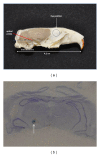
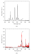
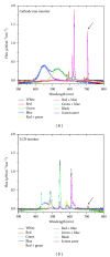
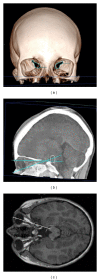
Recent data indicates that prolonged bright light exposure of rats induces production of neuromelanin and reduction of tyrosine hydroxylase positive neurons in the substantia nigra. This effect was the result of direct light reaching the substantia nigra and not due to alteration of circadian rhythms. Here, we measured the spectrum of light reaching the substantia nigra in rats and analysed the pathway that light may take to reach this deep brain structure in humans. Wavelength range and light intensity, emitted from a fluorescent tube, were measured, using a stereotaxically implanted optical fibre in the rat mesencephalon. The hypothetical path of environmental light from the eye to the substantia nigra in humans was investigated by computed tomography and magnetic resonance imaging. Light with wavelengths greater than 600 nm reached the rat substantia nigra, with a peak at 709 nm. Eyes appear to be the gateway for light to the mesencephalon since covering the eyes with aluminum foil reduced light intensity by half. Using computed tomography and magnetic resonance imaging of a human head, we identified the eye and the superior orbital fissure as possible gateways for environmental light to reach the mesencephalon.
Figures


References
-
- Ferrari M, Mottola L, Quaresima V. Principles, techniques, and limitations of near infrared spectroscopy. Canadian Journal of Applied Physiology. 2004;29(4):463–487. - PubMed
-
- Pervaiz S, Olivo M. Art and science of photodynamic therapy. Clinical and Experimental Pharmacology and Physiology. 2006;33(5-6):551–556. - PubMed
-
- Krawiecki Z, Cysewska-Sobusiak A, Wiczynski G, Odon A. Modeling and measurements of light transmission through human tissues. Bulletin of the Polish Academy of Sciences. 2008;56(2):147–154.
-
- Ansari MA. The effects of tissue optical parameters and interface reflectivity on light diffusion in biological tissues. World Academy of Science, Engineering and Technology. 2011;60:1875–1878.
Publication types
MeSH terms
LinkOut - more resources
Full Text Sources
Other Literature Sources

