. 2013 Jun;9(4):299-237.
doi: 10.2174/1573398X0904140129125307.

Pleuroparenchymal Fibroelastosis: Its Clinical Characteristics

Affiliations

Pleuroparenchymal Fibroelastosis: Its Clinical Characteristics

Kentaro Watanabe. Curr Respir Med Rev. 2013 Jun.

Abstract

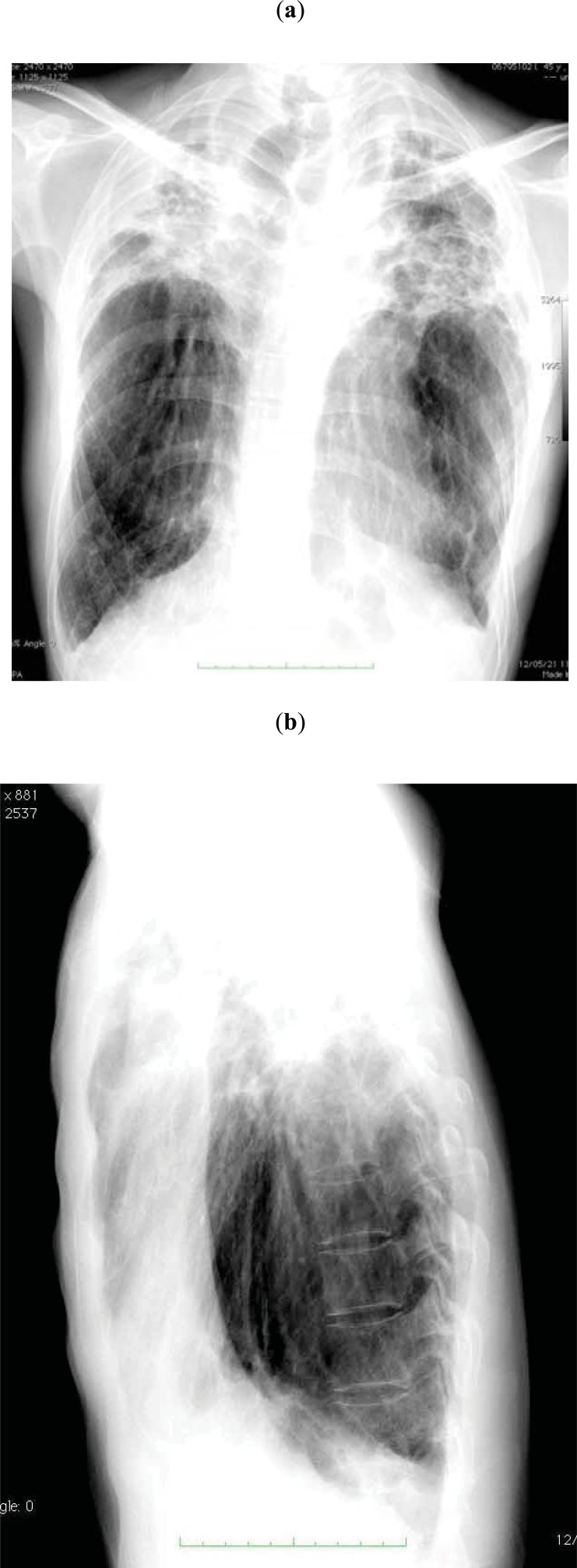
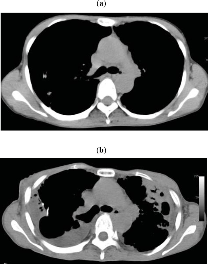
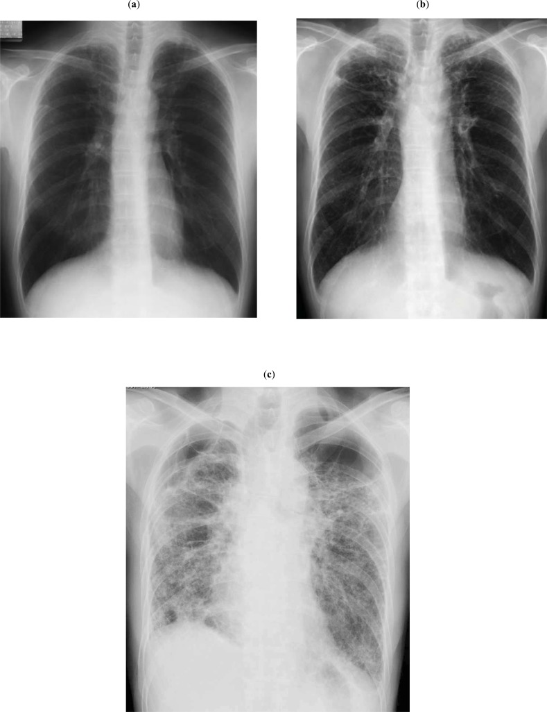
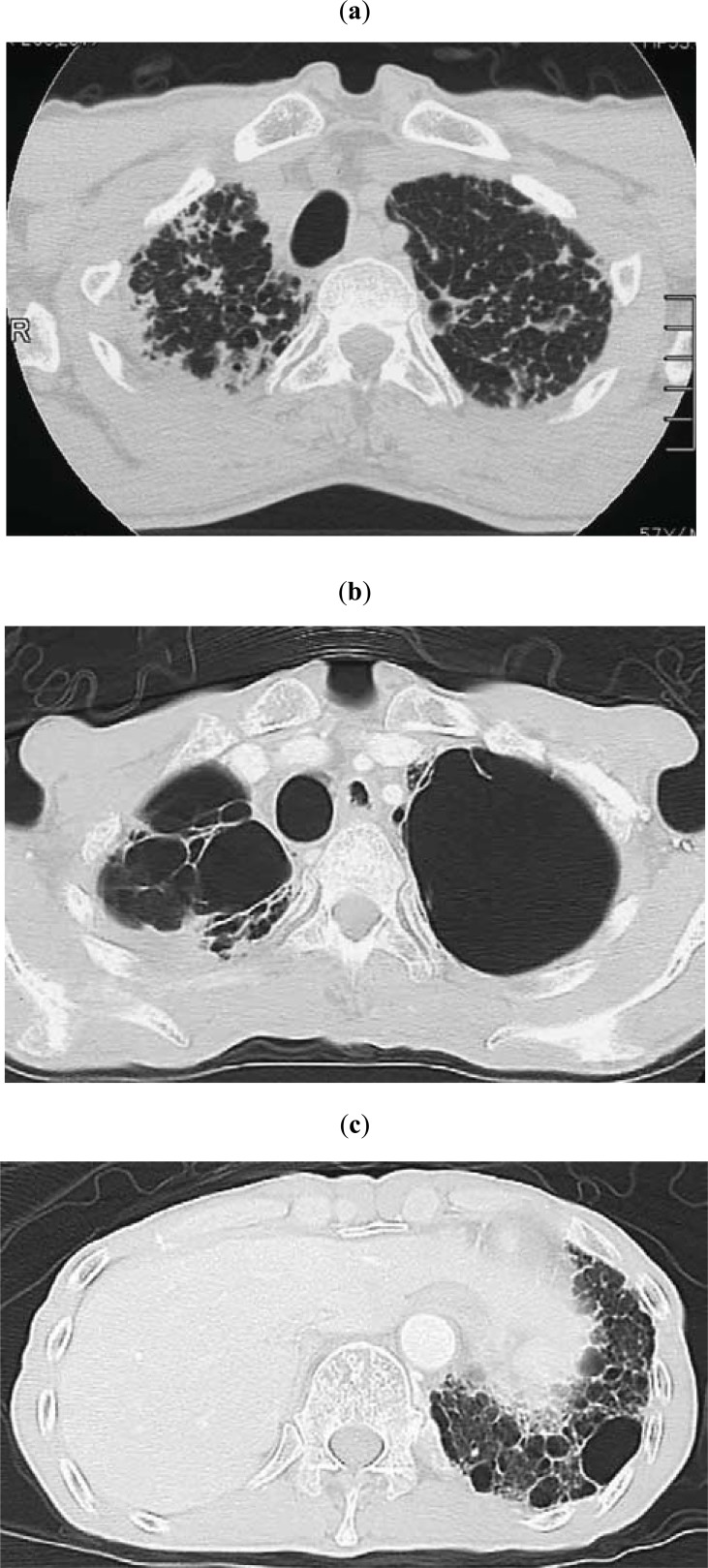
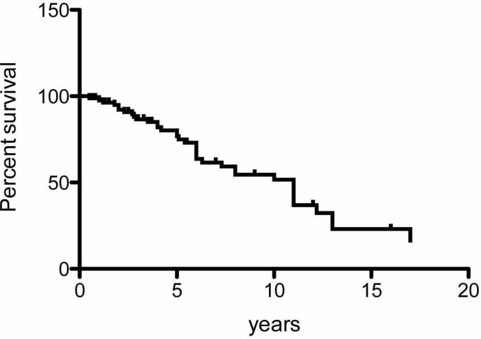
Pleuroparenchymal fibroelastosis (PPFE) is a rare pulmonary fibrosis that is clinically characterized by upperlobe predominant fibrosis. PPFE is a slowly progressive disorder and its first symptom is dyspnea or dry cough. Chest pain because of pneumothorax may be the first symptom in some patients. Patients with PPFE are slender with a flat rib cage or abnormally narrowed anterior-posterior thoracic dimension. Decreases in forced vital capacity, total lung capacity, and diffusing capacity are respiratory-function characteristics of PPFE, similar to those seen in idiopathic pulmonary fibrosis (IPF). The most remarkable difference in clinical features between PPFE and IPF is imaging findings, with upper-lobe-predominant lesions in PPFE and lower-lobe-predominant lesions in IPF.

Keywords: Pleuroparenchymal fibroelastosis (PPFE); pulmonary fibrosis (IPF).; pulmonary upper lobe fibrosis.


Figures

Fig. (1)
Fig. (1)
Chest radiographs of a PPFE patient (44-year-old man) showing a thin thoracic cage.
Fig. (2)
Fig. (2)
(a) Chest CT in a 48-year-old woman with PPFE. (b) Chest CT taken after 4 years. Both CT scans were sliced at the level of the 6th thoracic vertebra. The ratio of the anterior–posterior diameter of the thorax to the transverse diameter of the thorax became lower after 4 years of disease progression.
Fig. (3)
Fig. (3)
(a) Chest radiograph of a 50-year-old man with PPFE. At the early stage, the bilateral apical pleura appeared irregularly thickened; otherwise, they were almost normal. (b) Chest radiograph taken 4.7 years after the image shown in 3a. Reticular and nodular opacities appeared in the bilateral upper lung fields, and hilar opacities were further elevated. (c) Chest radiograph taken 9.7 years after the image shown in 3a. Fibrotic shadows extended to lower lung fields, and the diaphragm was elevated, with the loss of bilateral lung volume. Multiple bullae and large cysts appeared in the upper lung fields.
Fig. (4)
Fig. (4)
(Same patient as in Fig. 3) (a) Chest CT showing subpleural nodular or reticular opacities in the lung parenchyma at the apex. Interlobular septal thickening was associated (taken 7 years after the image shown in Fig. 3a). (b) Chest CT taken 2.8 years after the image shown in 4a. Multiple bullae and large cysts appeared in the upper lung fields. (c) Chest CT taken 2.8 years after the image shown in 4a. Multiple fibrocystic changes appear in the lower lobes.
Fig. (5)
Fig. (5)
A Kaplan-Meier survival curve using 85 patients from previous studies.

References

    1. Frankel SK, Cool CD, Lynch DA, Brown KK. Idiopathic pleuroparenchymal fibroelastosis.Description of a novel clinicopathologic Entity. . Chest. 2004;126:2007–13. - PubMed
    1. An Official American Thoracic Society/European Respiratory Society Statement: Update of the International multidisciplinary consensus classification of the idiopathic interstitial pneumonias. Amer J Respir Crit Care Med. 2013;188:733–48. - PMC - PubMed
    1. Davies D, Crowther JS, MacFarlane A. Idiopathic progressive pulmonary fibrosis. Thorax. 1975;30(3):316–25. - PMC - PubMed
    1. Davies D. Ankylosing spondylitis and lung fibrosis. Q J Med. 1972;41:395–417. - PubMed
    1. Clinicopathological Conference Undiagnosable lung disease demonstrated at the postgraduate medical school of London. Br Med J. 1962;1:1403–10. - PubMed

LinkOut - more resources