. 2014 Mar 18;53(10):1575-85.
doi: 10.1021/bi5000614. Epub 2014 Mar 6.

New insights into bacterial chemoreceptor array structure and assembly from electron cryotomography

Affiliations

New insights into bacterial chemoreceptor array structure and assembly from electron cryotomography

Ariane Briegel et al. Biochemistry. .

Erratum in

  • Biochemistry. 2014 Oct 21;53(41):6624

Abstract

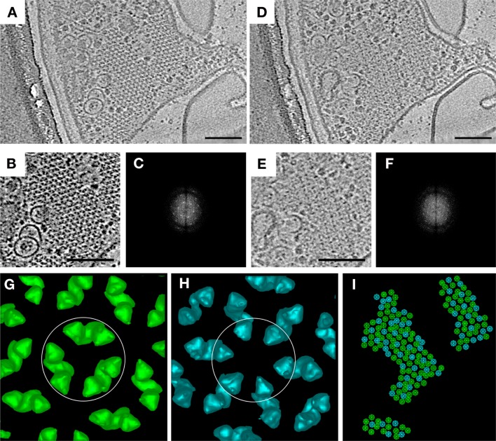
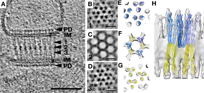
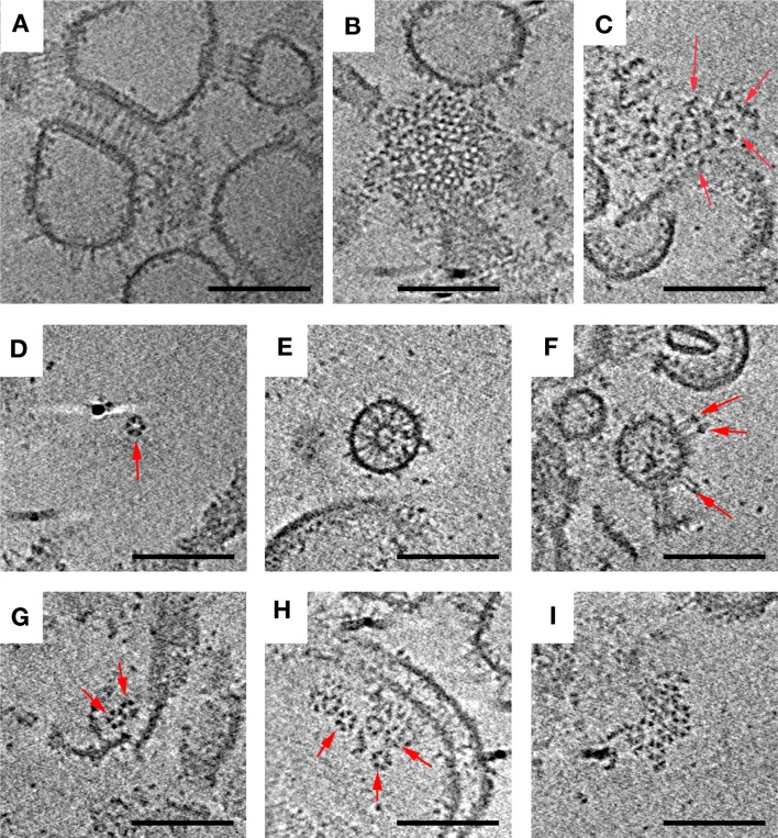
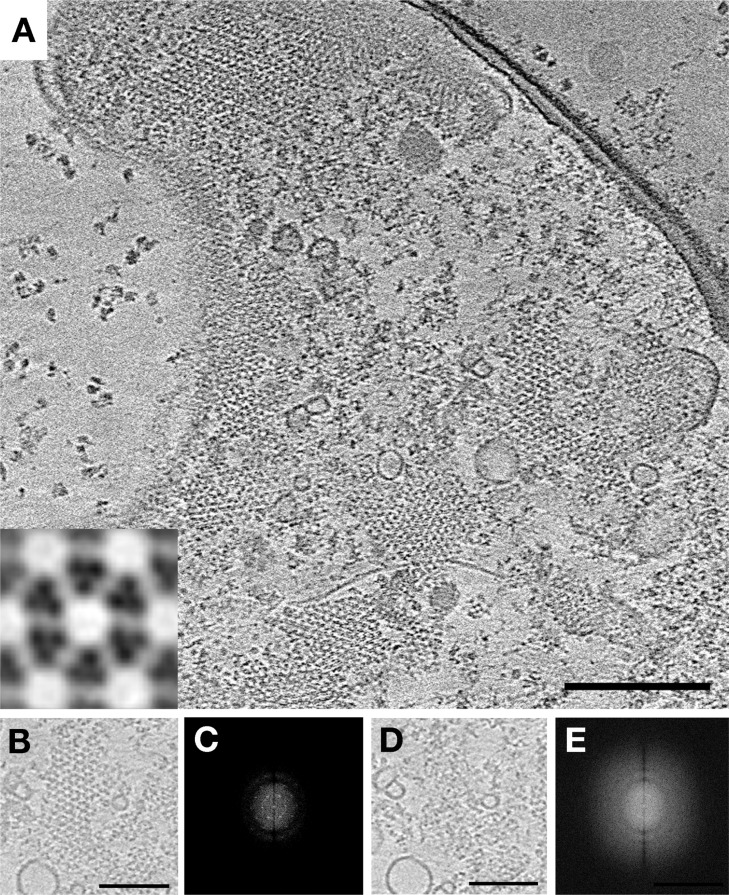
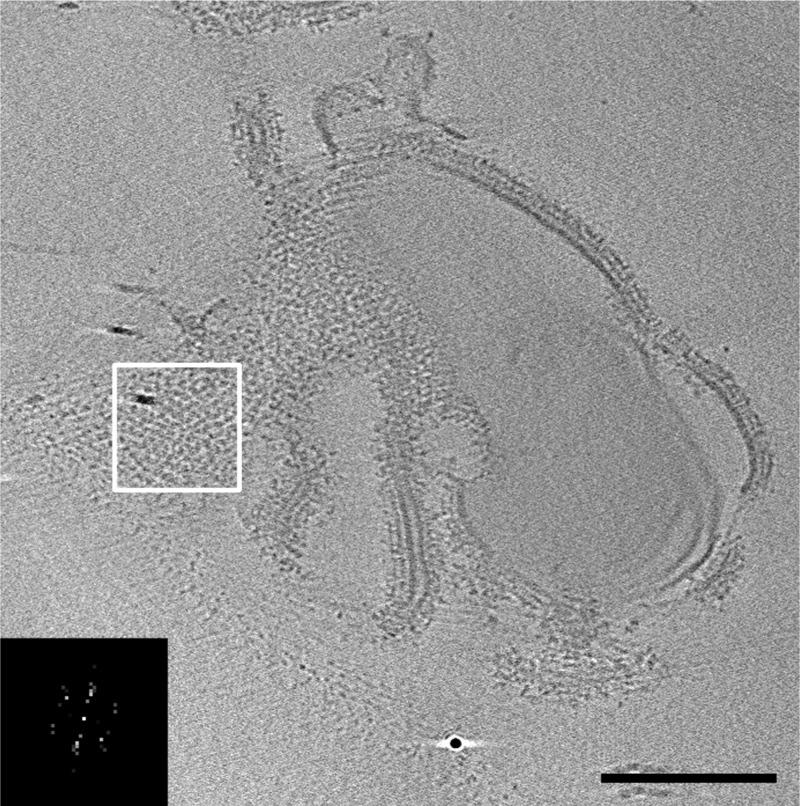
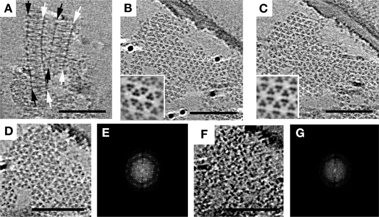
Bacterial chemoreceptors cluster in highly ordered, cooperative, extended arrays with a conserved architecture, but the principles that govern array assembly remain unclear. Here we show images of cellular arrays as well as selected chemoreceptor complexes reconstituted in vitro that reveal new principles of array structure and assembly. First, in every case, receptors clustered in a trimers-of-dimers configuration, suggesting this is a highly favored fundamental building block. Second, these trimers-of-receptor dimers exhibited great versatility in the kinds of contacts they formed with each other and with other components of the signaling pathway, although only one architectural type occurred in native arrays. Third, the membrane, while it likely accelerates the formation of arrays, was neither necessary nor sufficient for lattice formation. Molecular crowding substituted for the stabilizing effect of the membrane and allowed cytoplasmic receptor fragments to form sandwiched lattices that strongly resemble the cytoplasmic chemoreceptor arrays found in some bacterial species. Finally, the effective determinant of array structure seemed to be CheA and CheW, which formed a "superlattice" of alternating CheA-filled and CheA-empty rings that linked receptor trimers-of-dimer units into their native hexagonal lattice. While concomitant overexpression of receptors, CheA, and CheW yielded arrays with native spacing, the CheA occupancy was lower and less ordered, suggesting that temporal and spatial coordination of gene expression driven by a single transcription factor may be vital for full order, or that array overgrowth may trigger a disassembly process. The results described here provide new insights into the assembly intermediates and assembly mechanism of this massive macromolecular complex.


Figures

Figure 1
Figure 1
Classification of E. coli chemoreceptor array hexagons reveals ordered CheA occupancy. (A) Tomographic slice through an array patch at the level of the chemoreceptors. (B) Cutout of the patch, and corresponding power spectrum (C), revealing hexagonal lattice. (D) Tomographic slice of the same array patch below the level of the chemoreceptors, showing CheA. (E) Cutout of the patch, and corresponding power spectrum (F), revealing ordered occupancy by CheA. (G–I) Classification by principal components analysis and k-means clustering of hexagons in the same array patch results in two distinct classes: hexagons linked by three CheA dimers (green symbols, subvolume average circled in panel G) and hexagons lacking CheA dimers (turquoise symbols, subvolume average circled in panel H). The organization of class averages is shown in panel I. Scale bars are 100 nm, and power spectra are not to scale.
Figure 2
Figure 2
Overexpression of Tsr without sufficient CheA and CheW results in zippers. (A) A side view of a receptor zipper reveals two layers of membrane-bound receptors interacting at their membrane-distal tips. PD denotes periplasmic domains and IM inner membrane leaflets. The scale bar is 50 nm. Arrows indicate relative positions of subvolume averages shown at the right in panels B–D. Scale bars are 10 nm. (E–H) Model of receptor density from the subvolume average and manually fitted Tsr crystal structure from ref (46) in top view (E–G, levels roughly corresponding to B–D, respectively) and side view (H), showing the arrangement of receptors. Blue and yellow colors indicate receptors of opposing orientation.
Figure 3
Figure 3
In vitro reconstitution of signaling complexes produces a variety of structures. Arrangements observed included receptor zippers with 9 nm center-to-center hexagonal spacing (side view, A; top view, B), loosely ordered aggregates (C), individual hexagons of six trimers of dimers (D), receptors oriented inward (E) and outward (F) from vesicles, linked hexagons (G), multiple unlinked hexagons (H), and the largest 12 nm hexagonal array patch observed (I). Arrows indicate structures of interest. Scale bars are 100 nm.
Figure 4
Figure 4
Co-overexpression of Tsr, CheA, and CheW restores WT array structure. (A) A tomographic slice of a lysed E. coli cell overexpressing the chemotaxis proteins Tsr-A413T, CheA, and CheW reveals extended well-ordered hexagonal arrays with 12 nm center-to-center spacing. The inset shows a higher-magnification subvolume average showing the top view of a single hexagon. (B) Array patch at the level of the receptors and the corresponding power spectrum (C). (D) Same array patch at the level of CheA and the corresponding power spectrum (E) showing a lack of order in the CheA arrangement. Scale bars are 100 nm, and power spectra are not to scale.
Figure 5
Figure 5
Addition of MCPs after CheA and CheW produces extended 12 nm arrays. Vesicle-mediated assembly of Tar-CF, CheA, and CheW leads to extended arrays, shown in a tomographic slice. The inset shows a power spectrum (not to scale) of the white-boxed region that shows the hexagonal order of the array, with a 12 nm center-to-center spacing. The scale bar is 200 nm.
Figure 6
Figure 6
E. coli Tar chemoreceptors lacking transmembrane regions form extended arrays in the presence of CheA, CheW, and molecular crowding agents. Tomographic slices showing extended arrays. (A) A side view reveals two parallel CheA and CheW base plates (arrows) spaced 31 nm apart. Top views of the chemoreceptors close to either base plate (B and C, corresponding to white and black arrows in A, respectively) reveal a well-ordered, hexagonal arrangement with a center-to-center spacing of 12 nm. Insets show enlarged subvolume averages. (D) Array patch at the level of the receptors and the corresponding power spectrum (E). (F) Same array patch at the level of CheA and the corresponding power spectrum (G), showing the lack of order. Scale bars are 100 nm, and power spectra are not to scale.
Figure 7
Figure 7
Model of array assembly. Schematic showing sequential assembly of the core functional unit (two trimers-of-receptor dimers, one CheA dimer, and two CheW monomers) forming from individual trimers-of-receptor dimers, and subsequently coalescing into individual hexagons, which in turn assemble into the extended superlattice. Empty hexagons without associated CheA are colored blue.

References

    1. Hazelbauer G. L.; Falke J. J.; Parkinson J. S. (2008) Bacterial chemoreceptors: High-performance signaling in networked arrays. Trends Biochem. Sci. 33, 9–19. - PMC - PubMed
    1. Liu J.; Hu B.; Morado D. R.; Jani S.; Manson M. D.; Margolin W. (2012) Molecular architecture of chemoreceptor arrays revealed by cryoelectron tomography of Escherichia coli minicells. Proc. Natl. Acad. Sci. U.S.A. 109, E1481–E1488. - PMC - PubMed
    1. Briegel A.; Li X.; Bilwes A. M.; Hughes K. T.; Jensen G. J.; Crane B. R. (2012) Bacterial chemoreceptor arrays are hexagonally packed trimers of receptor dimers networked by rings of kinase and coupling proteins. Proc. Natl. Acad. Sci. U.S.A. 109, 3766–3771. - PMC - PubMed
    1. Briegel A.; Ortega D. R.; Tocheva E. I.; Wuichet K.; Li Z.; Chen S.; Müller A.; Iancu C. V.; Murphy G. E.; Dobro M. J.; Zhulin I. B.; Jensen G. J. (2009) Universal architecture of bacterial chemoreceptor arrays. Proc. Natl. Acad. Sci. U.S.A. 106, 17181–17186. - PMC - PubMed
    1. Briegel A.; Beeby M.; Thanbichler M.; Jensen G. J. (2011) Activated chemoreceptor arrays remain intact and hexagonally packed. Mol. Microbiol. 82, 748–757. - PMC - PubMed

Publication types

MeSH terms

LinkOut - more resources