. 2014 Mar 3;11(1):5.
doi: 10.1186/2045-8118-11-5.

Diverse arachnoid cyst morphology indicates different pathophysiological origins

Affiliations

Diverse arachnoid cyst morphology indicates different pathophysiological origins

Katrin Rabiei et al. Fluids Barriers CNS. .

Abstract

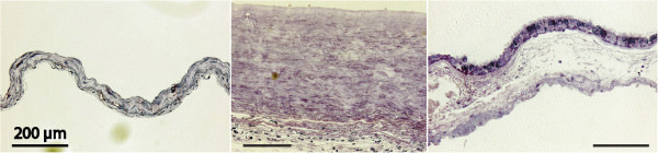
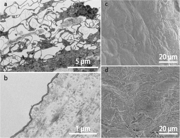
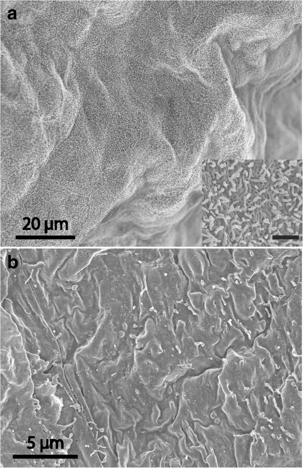
Background: There are few, limited, and to some extent contradictory, reports on the cellular and subcellular morphology of arachnoid cysts. In the literature cyst membranes are described as similar to, or as vastly different from, normal arachnoid membranes.

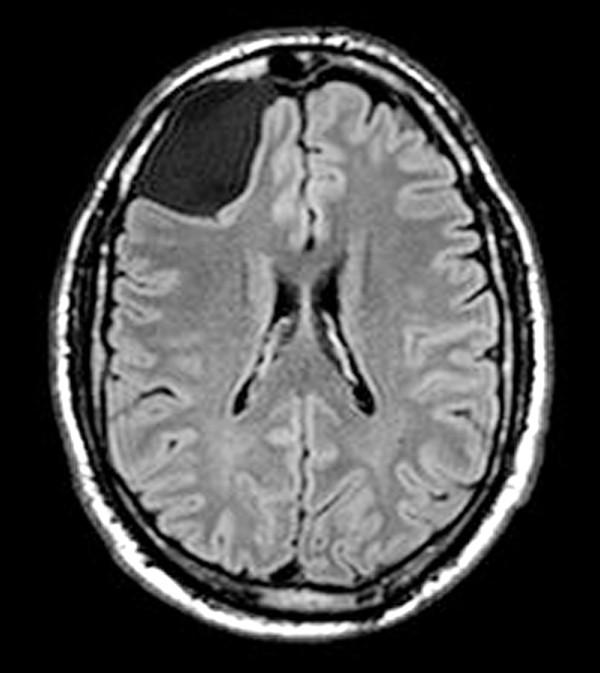
Methods: This paper reports electron microscopic analyses of symptomatic cysts from 24 patients (12 males and 12 females; age 10-79), that underwent fenestration surgery. Fourteen cysts were located in the middle cranial fossa (temporal), one in the interpeduncular cistern, five in the posterior fossa, and four were overlying the frontal cortex.

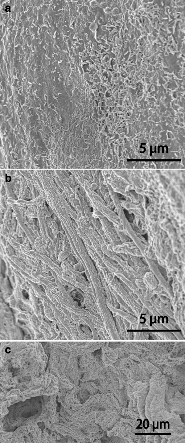
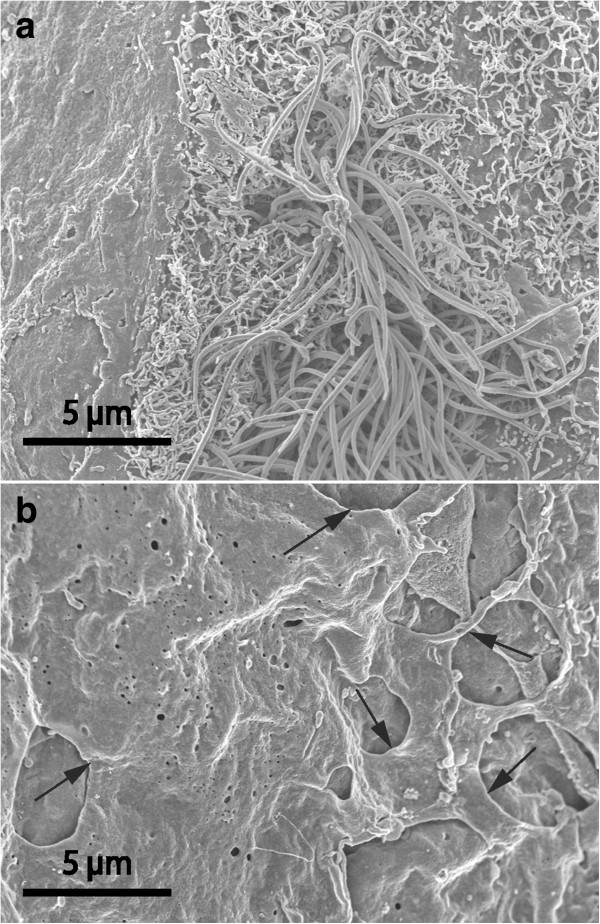
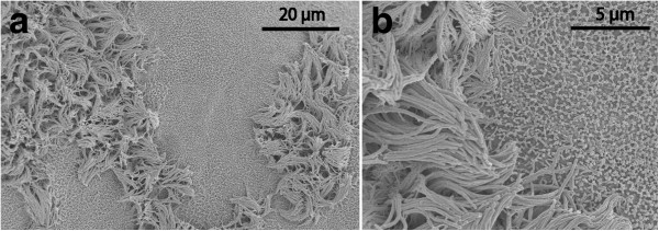
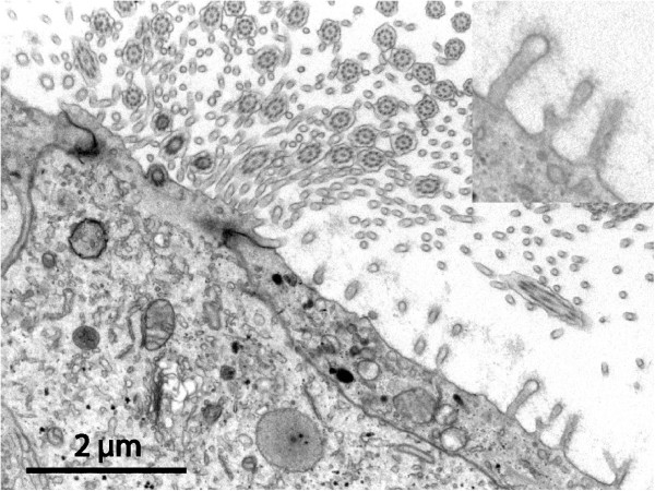
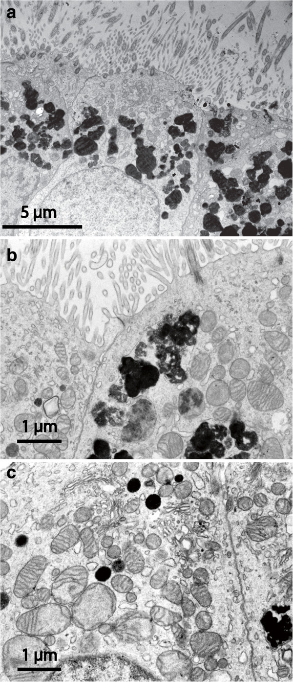
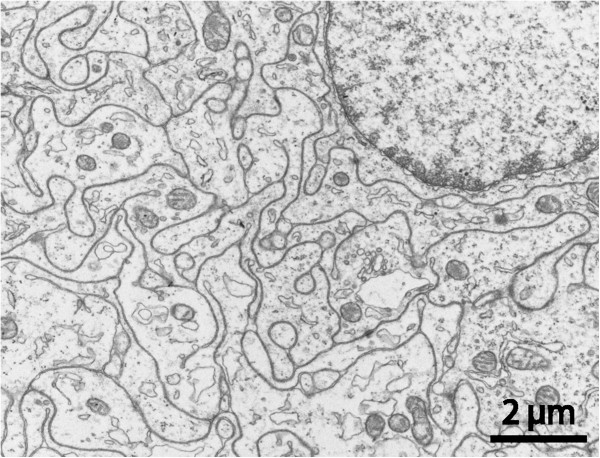
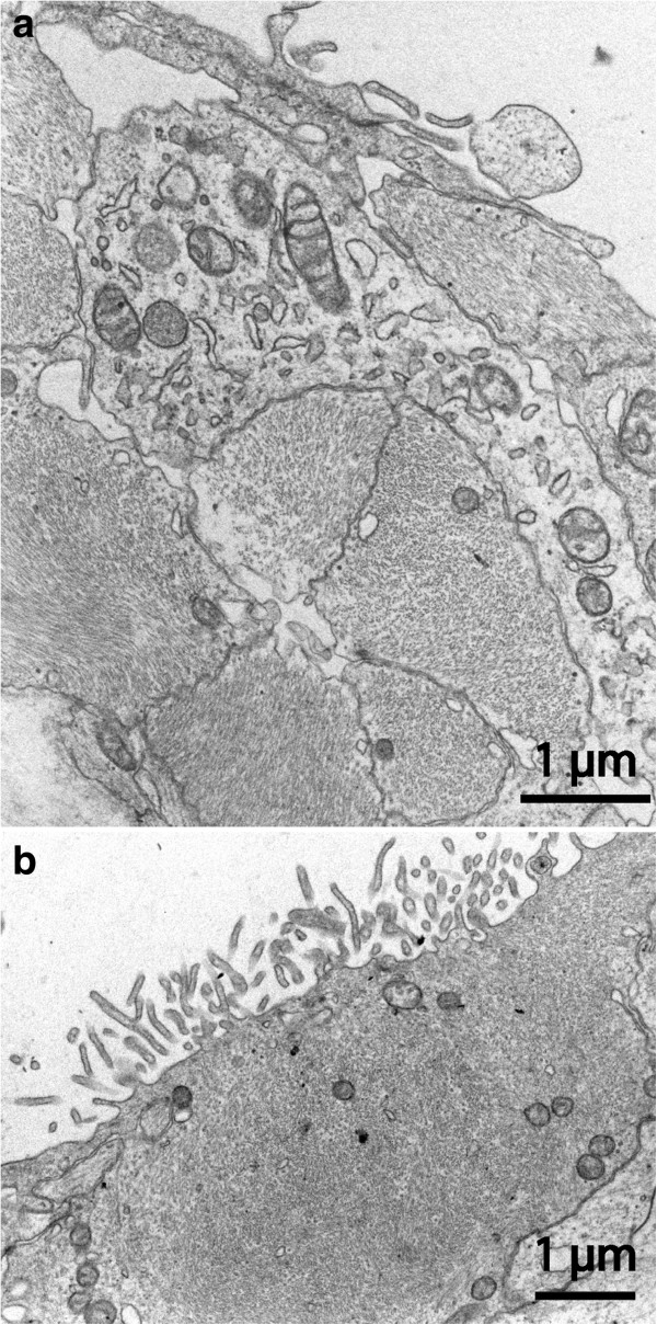
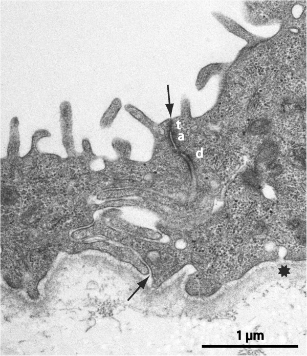
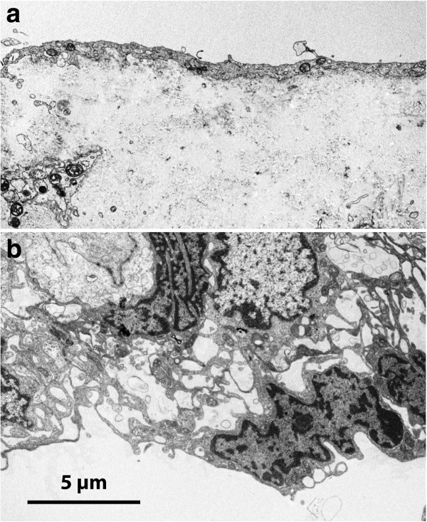
Results: Microscopic findings confirmed the diverse nature of this clinical condition. Twelve cyst walls resembled normal arachnoid, four had a conspicuous core of dense fibrous tissue with a simple epithelial lining, and the remaining aberrant cysts exhibited non-arachnoid luminal epithelia with plentiful microvilli and/or cilia, and also nervous tissue components in the wall. The possible identity and origin of various cyst types are discussed. We hypothesize that cysts are formed mostly at an early stage of embryonic development, as a teratological event.

Conclusions: Cysts with various epithelial linings and extracellular components most likely have different barrier properties and fluid turnover characteristics. Further studies are needed to elucidate relations between cyst morphology, fluid composition, pathogenesis, and clinical behaviour including growth rate and relapse tendency.


Figures

Figure 1
Figure 1
T1-weighted MRI image. A large arachnoid cyst deforms right frontal lobe; outer membrane of cyst is faintly visible. This patient presented with headache, vertigo, and seizures. The cyst wall consisted of respiratory-like epithelium.
Figure 2
Figure 2
Light microscopic overviews of 1 μm sections of cyst walls. Left to right: arachnoid, fibrous, and aberrant types, the latter with ciliated epithelium. Cyst lumen top, dural side below, Richardson’s stain, all scale bars = 200 μm.
Figure 3
Figure 3
EM findings on arachnoid and fibrous cysts. TEM of dural aspects of arachnoid cyst (a) and fibrous cyst (b); SEM of luminal surface of arachnoid cyst (c) and of fibrous cyst (d). A typical arachnoid organisation is seen in (a) with highly complex cellular extensions in whorls encircling wide intercellular spaces. In (b) only a single extremely attenuated cell covers a matrix rich in collagen and ground substance. In (c) and (d) scattered microvilli are seen as bright spots. The luminal surface epithelium is smooth in (c), more irregular with some overlapping or desquamating cells in (d).
Figure 4
Figure 4
Aberrant-type cyst. SEM micrographs of cyst with uniform appearance at both luminal (a) and dural surfaces (b). Luminal surface with dense distribution of short microvilli (see insert; bar 1 μm). The dural-aspect cells appear to be arranged in a tile-like manner and carry few microvilli.
Figure 5
Figure 5
Aberrant-type cysts. A comparison between luminal (a) and dural (b) cyst wall in a case where the SEM architecture of the dural side was particularly complex. Luminal cells (a) form a smooth surface with intermediate density of microvilli. Note elongated strands of cellular irregularities giving an uneven webbed dural surface (b). In comparison, (c) depicts an isolated sample where connective tissue filament bundles were exposed in the absence of covering cells on the luminal surface.
Figure 6
Figure 6
Aberrant-type cyst, SEM images. On luminal aspect (a) isolated ciliated cells were observed, here a microvillus-rich cells with a central tuft of cilia is surrounded by a profile-poor flat luminal epithelium. The dural surface (b) displayed numerous fine-edged openings (arrows) formed by cellular extensions and connections.
Figure 7
Figure 7
Aberrant-type cyst, SEM recordings of luminal surface. The epithelium exhibits a mosaic of two cell types, one equipped with numerous cilia, the other with extensive microvilli (a). On detailed examination (b), the bundles of long cilia are mixed with slender cellular projections (upper and lower left corners).
Figure 8
Figure 8
Aberrant-type cyst luminal surface. TEM micrograph of apical region of a high ciliated epithelial cell surrounded by flat microvilli-carrying cells (same specimen as in Figure 7). The two cell types meet in well-developed junctions. The blunt microvilli in the epithelial cell to the right have a constant length ending in a bulb-like widening, and carry a conspicuous fuzzy coat covering their surface (insert). Note also that the ciliated cell in addition to the obvious cilia also possesses numerous long and slender cytoplasmic extensions (mostly cross-sectioned) with a smaller diameter than the microvilli of the neighbouring cell.
Figure 9
Figure 9
Aberrant-type cyst: TEM details of luminal ciliated epithelium. In (a) and (b) slender long apical microvilli are more numerous than cilia. In the cytoplasm numerous irregular vesicles are seen with dense osmophilic content at various degrees of packing. The same cytoplasmic region is rich in mitochondria. Epithelial cells are closely adherent with probable tight junction complexes next to cyst lumen. In (c) a number of spherical vesicles with electron dense homogenous content are interspersed with numerous mitochondria.
Figure 10
Figure 10
Aberrant-type cyst. TEM section from cyst wall with numerous tightly interlocking cell processes containing microtubules, filaments, scattered vesicles and some mitochondria, i.e. exhibiting neuropil morphology. Occasional myelinated fibers were encountered (not shown).
Figure 11
Figure 11
Aberrant-type cyst. TEM micrographs from the luminal region of a cyst showing tightly packed cell profiles where the cytoplasm was almost completely occupied with masses of intermediate filaments. In (a) an organelle-rich cell is squeezed between filament-rich cells in both epithelial and sub-epithelial position, (b) shows a filament-rich cell with brush border-like microvilli towards cyst lumen.
Figure 12
Figure 12
Fibrous-type cyst, TEM micrograph. Junctional region of monolayered luminal epithelium, same specimen as Figure 3d. A complex, meandering, intercellular cleft runs between arrows; lumen up. Tight (t) and adherens (a) junction zones and a desmosome (d) are indicated. There is a distinct, continuous basal lamina (star).
Figure 13
Figure 13
TEM of comparison arachnoid from a patient with Chiari type I malformation. On pial aspect (a) an attenuated single-layered epithelium covers a cell poor matrix; the cellular arrangement towards the dura (b) is in agreement with normal arachnoid with the superficial cells forming complicated extensions in a network manner (cf. Figure 3a).

Similar articles

Cited by

References

    1. Wester K. Peculiarities of intracranial arachnoid cysts: location, sidedness, and sex distribution in 126 consecutive patients. Neurosurgery. 1999;45:775–779. doi: 10.1097/00006123-199910000-00008. - DOI - PubMed
    1. Rengachary SS, Watanabe I. Ultrastructure and pathogenesis of intracranial arachnoid cysts. J Neuropathol Exp Neurol. 1981;40:61–83. doi: 10.1097/00005072-198101000-00007. - DOI - PubMed
    1. Rengachary SS, Watanabe I, Brackett CE. Pathogenesis of intracranial arachnoid cysts. Surg Neurol. 1978;9:139–144. - PubMed
    1. Starkman SP, Brown TC, Linell EA. Cerebral arachnoid cysts. J Neuropathol Exp Neurol. 1958;17:484–500. doi: 10.1097/00005072-195807000-00009. - DOI - PubMed
    1. Choi JU, Kim DS. Pathogenesis of arachnoid cyst: congenital or traumatic? Pediatr Neurosurg. 1998;29:260–266. doi: 10.1159/000028733. - DOI - PubMed

LinkOut - more resources