

Effect of the vascular endothelial growth factor expression level on angiopoietin-2-mediated nasopharyngeal carcinoma growth
- PMID: 24581323
- PMCID: PMC4015607
- DOI: 10.1186/2045-824X-6-4
Effect of the vascular endothelial growth factor expression level on angiopoietin-2-mediated nasopharyngeal carcinoma growth
Abstract
Background: The overexpression of angiopoietin-2 (Ang-2) has both pro-tumorigenic and anti-tumorigenic effects. However, the mechanisms of this protein's dual effects are poorly understood, and it remains unclear how Ang-2 cooperates with vascular endothelial growth factor (VEGF). In the current study, we investigated the effects of Ang-2 overexpression on nasopharyngeal carcinoma growth in the presence of different levels of VEGF.
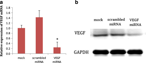
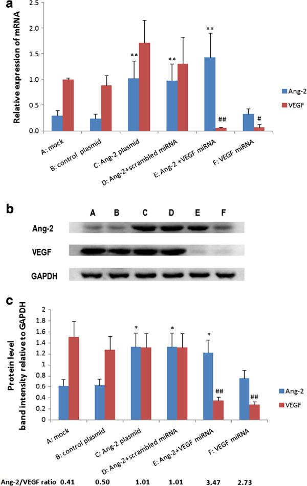
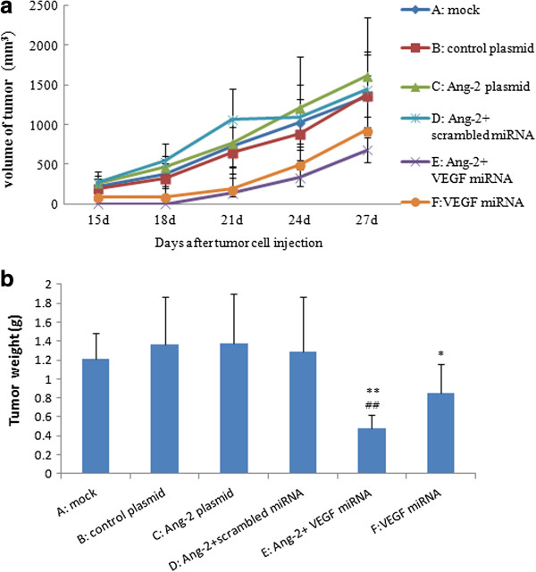
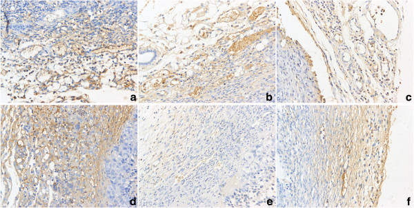
Methods: Ang-2 was introduced into the CNE2 cell line by liposome transfection, and the expression of endogenous VEGF was inhibited by microRNA-mediated RNA interference. CNE2 cells expressing varying levels of Ang-2 and VEGF were injected subcutaneously into the flanks of nude mice. Tumor growth was measured, and vessels from the harvested tumors were analyzed.
Results: The overexpression of Ang-2 had no obvious effect on CNE2 tumor growth in the presence of endogenous VEGF but significantly inhibited CNE2 tumor growth when the expression of endogenous VEGF was silenced, and the Ang-2/VEGF ratio is negatively correlated with tumor growth. Ang-2 overexpression decreased the percentage of α-SMA-positive cells around the tumor vessels but reduced the microvessel density only in the absence of VEGF.
Conclusions: Our results indicate that the effects of Ang-2 on nasopharyngeal carcinoma are highly dependent on the level of VEGF expression, Ang-2/VEGF ratio may offer a novel therapeutic approach for treating human cancer.
Figures

Similar articles
-
Tumor-derived vascular endothelial growth factor up-regulates angiopoietin-2 in host endothelium and destabilizes host vasculature, supporting angiogenesis in ovarian cancer.Cancer Res. 2003 Jun 15;63(12):3403-12. Cancer Res. 2003. PMID: 12810677
-
Angiopoietin-2 inhibits the growth of tongue carcinoma without affecting expression of vascular endothelial growth factor.Int J Oral Maxillofac Surg. 2011 Jun;40(6):628-32. doi: 10.1016/j.ijom.2010.11.005. Epub 2010 Dec 15. Int J Oral Maxillofac Surg. 2011. PMID: 21163623
-
Systemic overexpression of angiopoietin-2 promotes tumor microvessel regression and inhibits angiogenesis and tumor growth.Cancer Res. 2007 Apr 15;67(8):3835-44. doi: 10.1158/0008-5472.CAN-06-4056. Cancer Res. 2007. PMID: 17440098
-
Analysis of blood vessel maturation processes during cyclic ovarian angiogenesis.Lab Invest. 1998 Nov;78(11):1385-94. Lab Invest. 1998. PMID: 9840613
-
[Effects of norcantharidin on angiogenesis of human gallbladder carcinoma and its anti-angiogenic mechanisms].Zhonghua Yi Xue Za Zhi. 2006 Mar 14;86(10):693-9. Zhonghua Yi Xue Za Zhi. 2006. PMID: 16681930 Chinese.
Cited by
-
The effect of Setarud (IMOD(TM)) on angiogenesis in transplanted human ovarian tissue to nude mice.Iran J Reprod Med. 2015 Oct;13(10):605-14. Iran J Reprod Med. 2015. PMID: 26644788 Free PMC article.
-
Angiogenesis in nasopharyngeal carcinoma: insights, imaging, and therapeutic strategies.Front Oncol. 2024 May 28;14:1331064. doi: 10.3389/fonc.2024.1331064. eCollection 2024. Front Oncol. 2024. PMID: 38863627 Free PMC article. Review.
-
Potential suppressive effects of theophylline on human rectal cancer SW480 cells in vitro by inhibiting YKL-40 expression.Oncol Lett. 2018 May;15(5):7403-7408. doi: 10.3892/ol.2018.8220. Epub 2018 Mar 9. Oncol Lett. 2018. PMID: 29731892 Free PMC article.
-
Diagnostic and prognostic potential of serum angiopoietin-2 expression in human breast cancer.Int J Clin Exp Pathol. 2015 Jan 1;8(1):660-4. eCollection 2015. Int J Clin Exp Pathol. 2015. PMID: 25755760 Free PMC article.
-
Combining antiangiogenic therapy and radiation in nasopharyngeal carcinoma.Saudi Med J. 2015 Jun;36(6):659-64. doi: 10.15537/smj.2015.6.11460. Saudi Med J. 2015. PMID: 25987106 Free PMC article. Review.
References
-
- Imanishi Y, Hu B, Xiao G, Yao X, Cheng SY. Angiopoietin-2, an angiogenic regulator, promotes initial growth and survival of breast cancer metastases to the lung through the integrin-linked kinase (ILK)-AKT-B cell lymphoma 2 (Bcl-2) pathway. J Biol Chem. 2011;286:29249–29260. doi: 10.1074/jbc.M111.235689. - DOI - PMC - PubMed
-
- Leow CC, Coffman K, Inigo I, Breen S, Czapiga M, Soukharev S, Gingles N, Peterson N, Fazenbaker C, Woods R, Jallal B, Ricketts SA, Lavallee T, Coats S, Chang Y. MEDI3617, a human anti-angiopoietin 2 monoclonal antibody, inhibits angiogenesis and tumor growth in human tumor xenograft models. Int J Oncol. 2012;40:1321–1330. - PubMed
LinkOut - more resources
Full Text Sources
Other Literature Sources
Research Materials
Miscellaneous
