. 2014 Feb 27;156(5):907-19.
doi: 10.1016/j.cell.2014.01.042.

Stable C0T-1 repeat RNA is abundant and is associated with euchromatic interphase chromosomes

Affiliations

Stable C0T-1 repeat RNA is abundant and is associated with euchromatic interphase chromosomes

Lisa L Hall et al. Cell. .

Abstract

Recent studies recognize a vast diversity of noncoding RNAs with largely unknown functions, but few have examined interspersed repeat sequences, which constitute almost half our genome. RNA hybridization in situ using C0T-1 (highly repeated) DNA probes detects surprisingly abundant euchromatin-associated RNA comprised predominantly of repeat sequences (C0T-1 RNA), including LINE-1. C0T-1-hybridizing RNA strictly localizes to the interphase chromosome territory in cis and remains stably associated with the chromosome territory following prolonged transcriptional inhibition. The C0T-1 RNA territory resists mechanical disruption and fractionates with the nonchromatin scaffold but can be experimentally released. Loss of repeat-rich, stable nuclear RNAs from euchromatin corresponds to aberrant chromatin distribution and condensation. C0T-1 RNA has several properties similar to XIST chromosomal RNA but is excluded from chromatin condensed by XIST. These findings impact two "black boxes" of genome science: the poorly understood diversity of noncoding RNA and the unexplained abundance of repetitive elements.


Figures

Figure 1
Figure 1. CoT-1 RNA is expressed in all mammalian cells examined
All blue signals are DAPI DNA. CoT-1 RNA is expressed in 100% of interphase cells. e.g. A) HeLa (M=metaphase), B) NIH3T3. Inserts: CoT-1 RNA exclusion from chromocenters (inset arrows) of indicated cell (arrow). C) Human ES cells and D) frozen human tissue sections. D–E) Linescan across nuclei shows CoT-1 RNA absence from peripheral heterochromatic compartment (yellow zones). F–G) XIST RNA (arrow) paints the inactive X chr in female cells, while CoT-1 RNA is silenced. (See Fig S1 & Table S1). Scale bar 5µm.
Figure 2
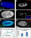
Figure 2. CoT-1 RNA appears highly abundant
A) XIST RNA paints interphase X-chromosome territory. Scale bar 5µm. B) The DAPI dense Barr Body (arrow) defines the inactive X-chromosome painted by XIST RNA. C) Both CoT-1 and XIST RNAs are detected simultaneously with the same fluorochrome. D) Linescan (white line in C) through the Barr body (arrow) illustrates similar intensities for both RNAs (green). E–H) CoT-1 RNA is more abundant than rRNA. All images are the same exposure. E) CoT-1 RNA alone, F) nucleolar 18S rRNA alone (Insert is same cell with DAPI DNA included), G) CoT-1 RNA and 18S rRNA detected with same fluorochrome. H) Average total intensities were measured for each RNA signal (n=40).
Figure 3
Figure 3. CoT-1 RNA localizes to the chromosome similar to XIST RNA
A) XIST RNA is strictly localized to the inactive chromosome in interphase. B) Human CoT-1 RNA is also strictly localized to human Chr4 in all interphase hybrid cells. Both RNAs are released at Mitosis (M) (arrows). C–D) Excised introns do not localize to chromatin, and drift away from their transcription sites. Transcription foci denoted by arrows. E) CoT-1 DNA identifies human Chr4 in hybrid cells painted by CoT-1 RNA (channels separated at right). Scale bar 5µm. F) A linescan (white line in E) shows the RNA has a sharp border at the edge of the chromosome territory. G) 3D image of CoT-1 DNA and RNA on human Chr4 in a single hybrid nucleus, with side views. H) same image as G with DAPI DNA removed. I) CoT-1 DNA and RNA signals are colocalized. (See Fig S1 & S2 and Movies S1, S2 & S3).
Figure 4
Figure 4. The majority of RNA associated with interphase chromosomes is repeat RNA, which is released at mitosis and resynthesized in G1
A–C) XIST RNA is released at mitosis and re-synthesized in early G1d cells. Scale bar 5µm. D–F) CoT-1 RNA is also released at mitosis and re-synthesized in G1d. G) A Chr4 library probe effectively detects the unique single copy DNA sequences across the chromosome (with CoT-1 competition). H–I) The same Chr4 library probe detects only a weak RNA signal (arrow) over the Chr4 territory (with CoT-1 competition). J–K) More RNA is detected on the Chr4 territory when CoT-1 competition is removed. H–K are taken at the same exposure with standardized fluorescent beads (small round objects in images). (See Fig S2). Beads are 2.5µm.
Figure 5
Figure 5. CoT-1 RNA localization is very stable under transcriptional inhibition
A–B) CoT-1 RNA is re-synthesized early in G1d (arrows). C–H) Transcriptional inhibition using three inhibitors (C–H) prevents CoT-1 re-synthesis in 95–100% of G1d cells (arrows), but doesn’t significantly affect CoT-1 RNA levels in 93–95% of interphase cells. I–L) COL1A1 transcription foci are detected in 95% of interphase and G1d cells (arrows). (I–J) COL1A1 RNA foci are eliminated in 82% of cells after 5 hours in DRB, but only G1d cells (arrows) also lack CoT-1 RNA. (See Fig S2 & S3). All scale bars are 5µm.
Figure 6
Figure 6. RNA from the 3’ end of L1 is a large component of the CoT-1 RNA signal
L1 ORF2 RNA signal (A–B) is ~4X brighter than the double stranded L1 ORF2 DNA signal (C–D), while the L1 ORF1 RNA signal (E–F) is ~0.2X the DNA signal (G–H). I) L1 RNA/DNA signals were quantified using digital fluorimetry. J) qPCR confirms L1 ORF2 RNA is ~5–7X more expressed than ORF1 RNA. K) Alu RNA is ~3–4X dimmer than its DNA signal quantified by digital fluorimetry (See Fig S5 & S6).
Figure 7
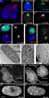
Figure 7. CoT-1 RNA associates with the nuclear scaffold and Cot-1 RNA loss is coincident with chromatin collapse
A) CoT-1 RNA remains bound to human Chr4 in ruptured nuclei. B–C) CoT-1 RNA is localized in control cells, and D–E) remains localized after removal of ~95% of DNA and histones (in 100% of interphase cells). Exposure times are equal for both images. F–G) CoT-1 RNA remains localized in 88% of cells containing wt SAF-A-GFP, while 80% of cells transfected with C280-GFP mutant (H–I) release CoT-1 RNA. Images include neighboring untransfected cells. J–K) Chromatin collapse seen by EM in transcriptionally inhibited G1d cells compared to similarly inhibited interphase cells (L). M) Transcriptionally inhibited interphase cells retain stable CoT-1 RNA, while post-mitotic cells do not. N) Chromatin does not collapse in interphase cells, but more compacted post-mitotic cells were seen in treated (86%) versus control (12%) samples. O–P) Interphase chromatin collapses when unfixed cells are treated with RNase. Scale bars are 5µm except for EM images which are 2µm.

Comment in

Similar articles

Cited by

References

    1. Aalto AP, Pasquinelli AE. Small non-coding RNAs mount a silent revolution in gene expression. Current opinion in cell biology. 2012;24:333–340. - PMC - PubMed
    1. Bensaude O. Inhibiting eukaryotic transcription: Which compound to choose? How to evaluate its activity? Transcription. 2011;2:103–108. - PMC - PubMed
    1. Britten RJ, Kohne DE. Repeated sequences in DNA: Hundreds of thousands of copies of DNA sequences have been incorporated into the genomes of higher organisms. Science. 1968;161:529–540. - PubMed
    1. Bynum JW, Volkin E. Chromatin-associated RNA: differential extraction and characterization. Biochimica et biophysica acta. 1980;607:304–318. - PubMed
    1. Chaumeil J, Le Baccon P, Wutz A, Heard E. A novel role for Xist RNA in the formation of a repressive nuclear compartment into which genes are recruited when silenced. Genes Dev. 2006;20:2223–2237. - PMC - PubMed

Publication types