Review
. 2014 Feb:17:17-23.
doi: 10.1016/j.mib.2013.10.007. Epub 2013 Dec 5.

Macrophages clean up: efferocytosis and microbial control

Affiliations
Review

Macrophages clean up: efferocytosis and microbial control

Constance J Martin et al. Curr Opin Microbiol. 2014 Feb.

Abstract

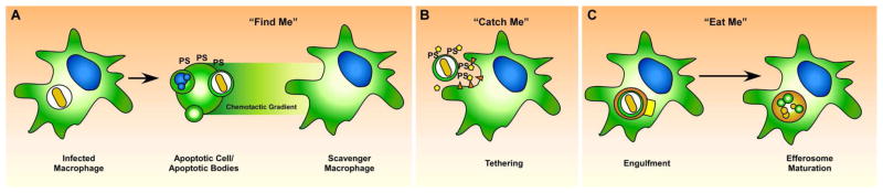
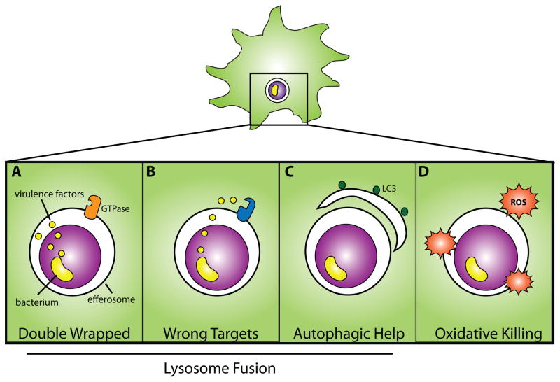
Phagocytic leukocytes, predominantly macrophages, not only ingest and destroy invading pathogens, but are charged with clearing dead and dying host cells. The process of engulfing apoptotic cells is called efferocytosis and has long been appreciated for its role in the resolution of inflammation. New evidence is emerging that efferocytosis represents a double-edged sword in microbial immunity. Although efferocytosis of influenza and Mycobacterium tuberculosis-infected cells results in pathogen destruction, efferocytosis of Leishmania-infected neutrophils may promote infection. Understanding how macrophages, dendritic cells (DC) and neutrophils process pathogens encased within a dying cell could lead to the development of novel therapeutics that simultaneously suppress inflammation and promote pathogen clearance.


Figures

Figure 1
Figure 1. Efferocytosis is a distinct process from phagocytosis
In phagocytosis, increased levels of RhoA result in actin polymerization and stress fiber formation, allowing receptor-mediated uptake of microbes. Fusion of the actin projections leads to internalization of the bacteria in a phagosome, which undergoes stepwise maturation, leading to degradation of the pathogen. This process can lead to both tissue sterilization and antigen presentation. In contrast, efferocytosis results from increased levels of Rac1, causing formation of lamellipodia and membrane ruffles upon actin polymerization, leading to the internalization of infected and ‘double-wrapped’ apoptotic bodies in an efferosome. From here, the ‘double-wrapped’ pathogen can be destroyed if the efferosome undergoes acidification, similar to the process of phagolysosome fusion. However, some pathogens appear to be able to use the process of efferocytosis to diseminate and disperse.
Figure 2
Figure 2. Efferocytosis involves the regulated uptake and degradation of apoptotic bodies
When an infected cell undergoes apoptosis (A), the pathogen and its antigens are presumably packaged along with other intracellular contents into apoptotic bodies which have intact cellular membranes. During apoptosis, the dying cell produces “Find Me” signals, such as chemokines that recruit macrophages and other phagocytic cells to the site of the dying cell, along with the accumulation of of phosphatidylserine (PS) on the exofacial leaflet. In the “Catch Me” phase, recruited macrophages express specific receptors and bridge molecules that bind to PS or other distinct ligands on the apoptotic cell (B). Binding between the apoptotic cell and the scavenger macrophage activates Rac1, leading to actin reorganization and production of projections that surround the infected apoptotic cell, ulimately surrounding and engulfing the apoptotic cell in the efferosome (C). The efferosome undergoes step-wise maturation that includes fusion with lysosomes and endosomes, leading to rapid degradation of both the apoptotic cell and the intracellular pathogen.
Figure 3
Figure 3. Four Proposed Mechanisms of Efferocytic Control
Following efferocytosis of an infected apoptotic body there are 4 potential mechanisms that contribute to microbial killing. (A) A microbe’s secreted virulence factors, which typically penetrate the phagosome, are trapped behind additional membrane from the apoptotic cell; consequently, they cannot reach their host targets. As it has been proposed that the machinery involved in efferosome maturation is different than phagosome maturation, (B), microbial virulence factors are ineffective at inhibiting efferosome maturation. Following efferocytosis, autophagic machinery can be recruited to the nascent efferosome (C). A, B, and C all ultimately allow lysosome fusion. In (D), reactive oxygen species generated in response to efferocytosis indirectly kills the pathogen.
Figure 4
Figure 4. Efferocytosis is one mechanism that the host can use to control infection
Macrophages autonomous control (A) frequently happens with low virulence or extracellular pathogens following phagocytosis. For pathogens that are more adapted to the intracellular environment of the macrophage, the macrophage needs help and collaborates with T cells (B). T cell recognition of the macrophage leads to the elaboration of T cell cytokines such as IFNγ and TNF, which activate the macrophage and enhance phagolysomal fusion but also stimulates other antimicrobial pathways, which augments the ability of the macrophage to kill intracellular bacteria. Some pathogens can overwhelm macrophage defenses. Under these conditions, the macrophage may die by apoptosis (C) if it is recognized by CTL; alternately, cell intrinsic factors may trigger an apoptotic death. Death by apoptosis ultimately allows for efferocytosis.

References

    1. Neefjes J, Jongsma ML, Paul P, Bakke O. Towards a systems understanding of MHC class I and MHC class II antigen presentation. Nat Rev Immunol. 2011;11 :823–836. - PubMed
    1. Savill J, Fadok V, Henson P, Haslett C. Phagocyte recognition of cells undergoing apoptosis. Immunol Today. 1993;14:131–136. - PubMed
    1. Taylor RC, Cullen SP, Martin SJ. Apoptosis: controlled demolition at the cellular level. Nat Rev Mol Cell Biol. 2008;9:231–241. - PubMed
    1. Rodriguez-Manzanet R, Sanjuan MA, Wu HY, Quintana FJ, Xiao S, Anderson AC, Weiner HL, Green DR, Kuchroo VK. T and B cell hyperactivity and autoimmunity associated with niche-specific defects in apoptotic body clearance in TIM-4-deficient mice. Proc Natl Acad Sci U S A. 2010;107:8706–8711. - PMC - PubMed
    1. Jaumouille V, Grinstein S. Receptor mobility, the cytoskeleton, and particle binding during phagocytosis. Curr Opin Cell Biol. 2011;23:22–29. - PubMed

Publication types

LinkOut - more resources