

Review
doi: 10.1016/j.mib.2013.11.001.
Epub 2013 Dec 5.
Structural organisation of the type IV secretion systems
Affiliations
- PMID: 24581689
- PMCID: PMC3969286
- DOI: 10.1016/j.mib.2013.11.001
Item in Clipboard
Review
Structural organisation of the type IV secretion systems
Curr Opin Microbiol.
2014 Feb.
Abstract
Type IV secretion (T4S) systems are large dynamic nanomachines that transport DNAs and/or proteins through the membranes of bacteria. Because of their complexity and multi-protein organisation, T4S systems have been extremely challenging to study structurally. However in the past five years significant milestones have been achieved by X-ray crystallography and cryo-electron microscopy. This review describes some of the more recent advances: the structures of some of the protein components of the T4S systems and the complete core complex structure that was determined using electron microscopy.
Copyright © 2013 The Authors. Published by Elsevier Ltd.. All rights reserved.
Figures
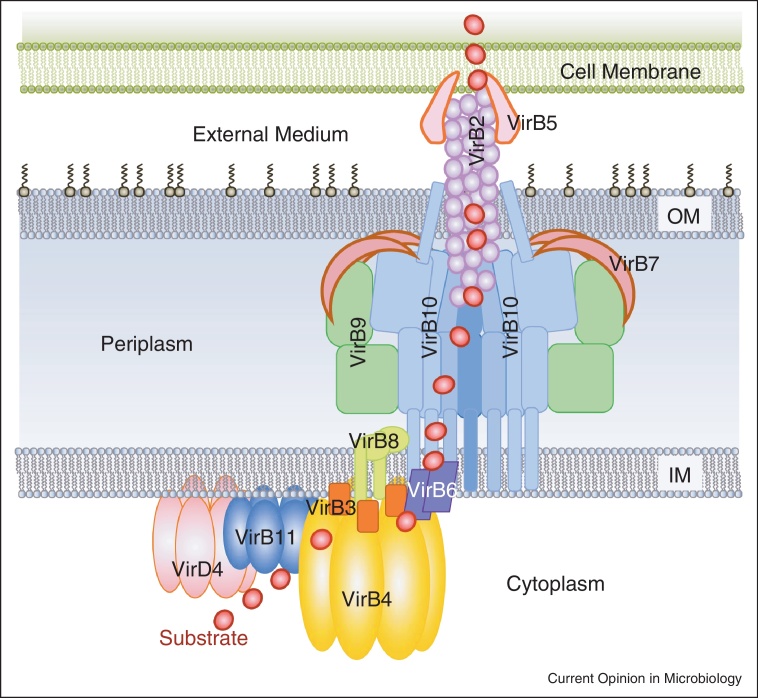
Overall organisation of the T4S system. VirD4 (in pink), VirB11 (in blue), VirB4 (in gold) ATPases, polytopic VirB6 (in purple), bitopic VirB8 (in light green) and VirB3 (in orange) form the cytoplasmic IM part of the complex. VirB7 (in brown), VirB9 (in green), and VirB10 (in blue) compose the periplasmic part of the secretion system. VirB2 and VirB5 constitute the outer part of the secretion system. Red dot indicates the path of the substrate through the machinery as established by Cascales and Christie [22•]. The stoichiometry of the various components in a native, fully assembled, T4S system is unknown.
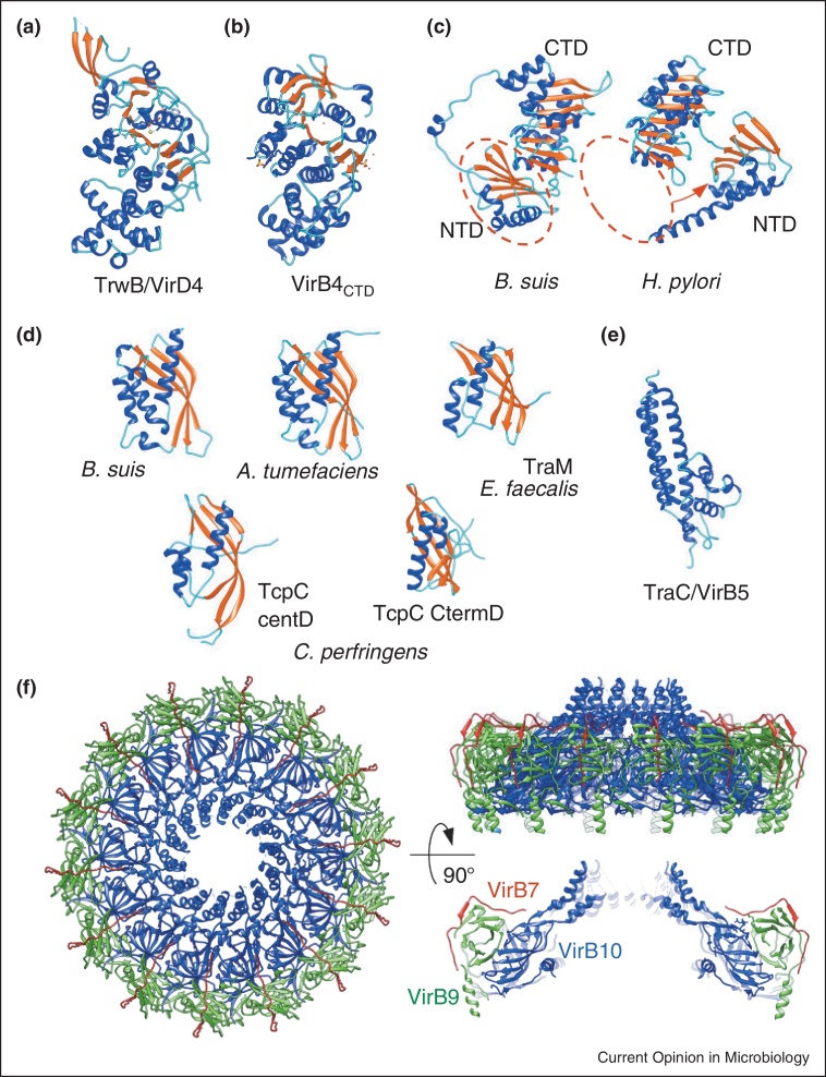
Structures of T4S system components or domains. (a) VirD4: structure of the soluble domain of TrwB. One subunit of the hexamer is shown (coils are shown in cyan, helices in blue, and strands in red). (b) VirB4: C-terminal domain of Thermoanaerobacter pseudethanolicus VirB4; (c) VirB11: crystal structures of B. suis VirB11 and H. pylori HP5025, CTD-C terminal domain, NTD-N terminal domain (shown in red dashed oval on B. suis VirB11). Red arrow indicates the shift of the NTD in H. pylori Vir11 compared to B. suis VirB11. (d) VirB8: crystal structure of the periplasmic domain (C-terminal domain) of VirB8 from B. suis (left panel), A. tumefaciens (middle panel), TraM214–322 protein (right panel); the central and C-terminal domains of the TcpC99–359 structure are shown in the bottom panel. (e) VirB5: crystal structure of TraC encoded by the E. coli conjugative plasmid pKM101. (f) Crystal structure of the CC's O-layer composed of VirB7 (in dark red), VirB9CT (in green) and VirB10CT (blue). All structures are shown in the ribbon representation.
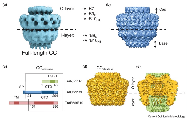
Electron microscopy of the T4S system. (a) Structure of the CC complex at 15 Å; (b) structure of the CC complex at 12 Å; (c) schematic illustration of the regions of TraN/VirB7, TraO/VirB9, and TraF/VirB10 present in the CCelastase complex. Domains corresponding to the VirB9 binding domain (B9BD), the signal peptide (SP), the N-terminal trans-membrane (TM) helix and the C-terminal domains (CTD) are shown in darker colours; (d) Cryo-EM structure of the CCelastase complex. (e) Cutaway view of the superposition of the difference map (in green) between the full length CC and CCelastase and the cryo-EM structure of the CCelastase complex (in gold).
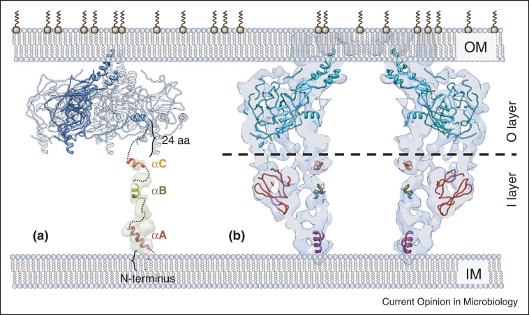
Schematic model for the full length organisation of the core complex. (a) Four TraF/VirB10CT subunits of the 14-mer present in the O-layer atomic structure are shown. One subunit is highlighted in blue. The density of one subunit column in the difference map (in light green) is shown with the tentative docking of the three TraF/VirB10NT α-helical regions. This subunit is located immediately below TraF/VirB10CT shown in dark blue. The connections are shown in dashed lines. (b) Central slice of the full length CC with fitted O-layer atomic structure (in cyan) and atomic models obtained for TraO/VirB9NT (in dark red) and the α-helices predicted in TraF/VirB10NT (shown as in (a)).
Similar articles
-
Structure of a bacterial type IV secretion core complex at subnanometre resolution.EMBO J. 2013 Apr 17;32(8):1195-204. doi: 10.1038/emboj.2013.58. Epub 2013 Mar 19. EMBO J. 2013. PMID: 23511972 Free PMC article.
-
Type IV secretion machinery: molecular architecture and function.Biochem Soc Trans. 2013 Feb 1;41(1):17-28. doi: 10.1042/BST20120332. Biochem Soc Trans. 2013. PMID: 23356253 Review.
-
Bacterial type IV secretion systems in human disease.Mol Microbiol. 2009 Jul;73(2):141-51. doi: 10.1111/j.1365-2958.2009.06751.x. Epub 2009 Jun 8. Mol Microbiol. 2009. PMID: 19508287 Free PMC article. Review.
-
Molecular architecture of bacterial type IV secretion systems.Trends Biochem Sci. 2010 Dec;35(12):691-8. doi: 10.1016/j.tibs.2010.06.002. Epub 2010 Jul 10. Trends Biochem Sci. 2010. PMID: 20621482 Review.
-
Cryo-EM structure of the bacteria-killing type IV secretion system core complex from Xanthomonas citri.Nat Microbiol. 2018 Dec;3(12):1429-1440. doi: 10.1038/s41564-018-0262-z. Epub 2018 Oct 22. Nat Microbiol. 2018. PMID: 30349081 Free PMC article.
Cited by
-
Architecture of the outer-membrane core complex from a conjugative type IV secretion system.Nat Commun. 2021 Nov 25;12(1):6834. doi: 10.1038/s41467-021-27178-8. Nat Commun. 2021. PMID: 34824240 Free PMC article.
-
Arabidopsis RETICULON-LIKE3 (RTNLB3) and RTNLB8 Participate in Agrobacterium-Mediated Plant Transformation.Int J Mol Sci. 2018 Feb 24;19(2):638. doi: 10.3390/ijms19020638. Int J Mol Sci. 2018. PMID: 29495267 Free PMC article.
-
Xanthomonas spp. Infecting Araceae and Araliaceae: Taxonomy, Phylogeny, and Potential Virulence Mechanisms.Biology (Basel). 2025 Jun 25;14(7):766. doi: 10.3390/biology14070766. Biology (Basel). 2025. PMID: 40723327 Free PMC article. Review.
-
Comparative Genome Analysis of Two Isolates of the Fish Pathogen Piscirickettsia salmonis from Different Hosts Reveals Major Differences in Virulence-Associated Secretion Systems.Genome Announc. 2014 Dec 18;2(6):e01219-14. doi: 10.1128/genomeA.01219-14. Genome Announc. 2014. PMID: 25523762 Free PMC article.
-
Draft genome sequence for virulent and avirulent strains of Xanthomonas arboricola isolated from Prunus spp. in Spain.Stand Genomic Sci. 2016 Jan 28;11:12. doi: 10.1186/s40793-016-0132-3. eCollection 2016. Stand Genomic Sci. 2016. PMID: 26823958 Free PMC article.
References
-
- Omori K., Idei A. Gram-negative bacterial ATP-binding cassette protein exporter family and diverse secretory proteins. J Biosci Bioeng. 2003;95:1–12. - PubMed
-
- Filloux A. The underlying mechanisms of type II protein secretion. Biochim Biophys Acta. 2004;1694:163–179. - PubMed
-
- Cornelis G.R., Van Gijsegem F. Assembly and function of type III secretory systems. Annu Rev Microbiol. 2000;54:735–774. - PubMed
Publication types
MeSH terms
Substances
Grants and funding
LinkOut - more resources
Full Text Sources
Other Literature Sources
Miscellaneous
