Review
. 2014 Apr;13(4):419-28.
doi: 10.1016/S1474-4422(14)70003-1. Epub 2014 Feb 27.

Outcome markers for clinical trials in cerebral amyloid angiopathy

Affiliations
Review

Outcome markers for clinical trials in cerebral amyloid angiopathy

Steven M Greenberg et al. Lancet Neurol. 2014 Apr.

Abstract

Efforts are underway for early-phase trials of candidate treatments for cerebral amyloid angiopathy, an untreatable cause of haemorrhagic stroke and vascular cognitive impairment. A major barrier to these trials is the absence of consensus on measurement of treatment effectiveness. A range of potential outcome markers for cerebral amyloid angiopathy can be measured against the ideal criteria of being clinically meaningful, closely representative of biological progression, efficient for small or short trials, reliably measurable, and cost effective. In practice, outcomes tend either to have high clinical salience but low statistical efficiency, and thus more applicability for late-phase studies, or greater statistical efficiency but more limited clinical meaning. The most statistically efficient markers might be those that are potentially reversible with treatment, although their clinical significance remains unproven. Many of the candidate outcomes for cerebral amyloid angiopathy trials are probably applicable also to other small-vessel brain diseases.


Conflict of interest statement

Conflicts of Interest: Massachusetts General Hospital, University of Calgary, Washington University/Barnes Jewish Hospital, Leiden University Medical Center, and Lille University Hospital have clinical research support agreements with Pfizer, sponsor of an ongoing trial for CAA. Dr. Schneider has received consulting fees or sat on paid advisory boards related to amyloid imaging for AVID radiopharmaceuticals, Eli Lilly Inc., and GE Healthcare. No other authors had conflicts of interest relevant to this manuscript.

Figures

Figure 1
Figure 1
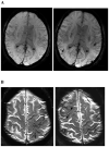
Examples of MRI markers. Panels A shows a 74 year old man with a total of 6 strictly lobar microbleeds on baseline T2*-weighted MRI (left-hand panel shows 1 of these) and 5 incident strictly lobar microbleeds on follow-up performed 3.4 years later (right-hand panel shows 3 of the incident lesions, arrows). Panels B show superficial siderosis on T2*-weighted MRI in a 69 year old man with probable CAA and predominantly left hemispheric siderosis at baseline (left) and incident right superior frontal siderosis on follow-up 1.5 years later (right, arrows). Panels C show growth of WMH on FLAIR images between baseline (left) and follow-up at 5 years (right) in an 89 year old man with probable CAA, old right occipital ICH, and progressive impairment of executive function but no ICH during the inter-scan interval. WMH volumes in the left (non-ICH) hemisphere were 18.5 cc at baseline and 23.9 cc at follow-up. Panels D shows a left frontal cortex focus of restricted diffusion (DWI image on left, absolute diffusion coefficient map on right) consistent with a small acute infarct in a 74 year old man with probable CAA and no other known vascular disease. Panel E shows sagittal FLAIR (left) and T1 (right) images obtained by 7T MRI of a 62 year old man with mild cognitive impairment. The lesions suggestive of cortical microinfacrts appear hyperintense on FLAIR and hypointense on T1 (arrows and insets for enlargement).
Figure 1
Figure 1
Examples of MRI markers. Panels A shows a 74 year old man with a total of 6 strictly lobar microbleeds on baseline T2*-weighted MRI (left-hand panel shows 1 of these) and 5 incident strictly lobar microbleeds on follow-up performed 3.4 years later (right-hand panel shows 3 of the incident lesions, arrows). Panels B show superficial siderosis on T2*-weighted MRI in a 69 year old man with probable CAA and predominantly left hemispheric siderosis at baseline (left) and incident right superior frontal siderosis on follow-up 1.5 years later (right, arrows). Panels C show growth of WMH on FLAIR images between baseline (left) and follow-up at 5 years (right) in an 89 year old man with probable CAA, old right occipital ICH, and progressive impairment of executive function but no ICH during the inter-scan interval. WMH volumes in the left (non-ICH) hemisphere were 18.5 cc at baseline and 23.9 cc at follow-up. Panels D shows a left frontal cortex focus of restricted diffusion (DWI image on left, absolute diffusion coefficient map on right) consistent with a small acute infarct in a 74 year old man with probable CAA and no other known vascular disease. Panel E shows sagittal FLAIR (left) and T1 (right) images obtained by 7T MRI of a 62 year old man with mild cognitive impairment. The lesions suggestive of cortical microinfacrts appear hyperintense on FLAIR and hypointense on T1 (arrows and insets for enlargement).
Figure 2
Figure 2
Serial functional MRI measurement of response to visual stimulation. Exemplar fMRI studies are shown from a 83 year old woman with probable CAA. fMRI with visual stimulation was performed at baseline (blue line, red error space) and again using the same scanner and protocol after 1 clinically asymptomatic year (green line, yellow error space). The blue and green solid lines represent the change from baseline BOLD signal averaged over 16 cycles of visual stimulation (on 20 sec, shaded region, then off 28 sec), with standard deviations of the response shown in red and yellow spaces and the trapezoidal model fits in solid and dashed black lines as described. The amplitude of the modeled peak response in this subject decreased from 1.06% at baseline to 0.80% at 1 year.

Comment in

References

    1. Viswanathan A, Greenberg SM. Cerebral amyloid angiopathy in the elderly. Ann Neurol. 2011;70(6):871–80. - PMC - PubMed
    1. Knudsen KA, Rosand J, Karluk D, Greenberg SM. Clinical diagnosis of cerebral amyloid angiopathy: Validation of the Boston Criteria. Neurology. 2001;56(4):537–9. - PubMed
    1. Pfizer. Study evaluating the safety, tolerability and efficacy of PF-04360365 in adults with probable cerebral amyloid angiopathy. ClinicalTrialsgov. 2013:NCT01821118. Available from: http://clinicaltrials.gov/ct2/show/NCT01821118?term=ponezumab&rank=5.
    1. Poon MT, Fonville AF, Al-Shahi Salman R. Long-term prognosis after intracerebral haemorrhage: systematic review and meta-analysis. Journal of neurology, neurosurgery, and psychiatry. 2013 Nov 21; doi: 10.1136/jnnp-2013-306476. Epub ahead of print. - DOI - PubMed
    1. Wardlaw JM, Smith EE, Biessels GJ, Cordonnier C, Fazekas F, Frayne R, et al. Neuroimaging standards for research into small vessel disease and its contribution to ageing and neurodegeneration. Lancet Neurol. 2013;12(8):822–38. - PMC - PubMed