Practice Guideline
. 2014 Apr;24(4):289-311.
doi: 10.1016/j.nmd.2013.12.011. Epub 2014 Jan 9.

Diagnostic approach to the congenital muscular dystrophies

Collaborators, Affiliations
Practice Guideline

Diagnostic approach to the congenital muscular dystrophies

Carsten G Bönnemann et al. Neuromuscul Disord. 2014 Apr.

Abstract

Congenital muscular dystrophies (CMDs) are early onset disorders of muscle with histological features suggesting a dystrophic process. The congenital muscular dystrophies as a group encompass great clinical and genetic heterogeneity so that achieving an accurate genetic diagnosis has become increasingly challenging, even in the age of next generation sequencing. In this document we review the diagnostic features, differential diagnostic considerations and available diagnostic tools for the various CMD subtypes and provide a systematic guide to the use of these resources for achieving an accurate molecular diagnosis. An International Committee on the Standard of Care for Congenital Muscular Dystrophies composed of experts on various aspects relevant to the CMDs performed a review of the available literature as well as of the unpublished expertise represented by the members of the committee and their contacts. This process was refined by two rounds of online surveys and followed by a three-day meeting at which the conclusions were presented and further refined. The combined consensus summarized in this document allows the physician to recognize the presence of a CMD in a child with weakness based on history, clinical examination, muscle biopsy results, and imaging. It will be helpful in suspecting a specific CMD subtype in order to prioritize testing to arrive at a final genetic diagnosis.

Keywords: Alpha-dystroglycan; Collagen VI; Congenital muscular dystrophy; Diagnostic guideline; Lamin A/C; Laminin alpha2; RYR1; SEPN1.


Conflict of interest statement

Authors declared that there is no conflict of interest.

Figures
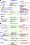
Fig. 1
Fig. 1
A–D: Differential diagnostic considerations for various clinical findings in infancy (A) and beyond infancy (B and C), as well as for various laboratory findings that may be available at the outset of the diagnostic encounter (D). Note: The most important tools in the clinical differential diagnosis are: EMG/NCV to diagnose neurogenic involvement, muscle biopsy, and selective biochemical and genetic testing. The differential diagnostic considerations are not exhaustive but highlight a few of the more relevant conditions to consider with a given clinical picture. To save space we are only using the gene/protein symbols to indicate specific diagnosis.
Fig. 1
Fig. 1
A–D: Differential diagnostic considerations for various clinical findings in infancy (A) and beyond infancy (B and C), as well as for various laboratory findings that may be available at the outset of the diagnostic encounter (D). Note: The most important tools in the clinical differential diagnosis are: EMG/NCV to diagnose neurogenic involvement, muscle biopsy, and selective biochemical and genetic testing. The differential diagnostic considerations are not exhaustive but highlight a few of the more relevant conditions to consider with a given clinical picture. To save space we are only using the gene/protein symbols to indicate specific diagnosis.
Fig. 2
Fig. 2
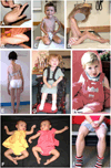
A: Hand of a patient with COL6-RD. Note the significant hyperlaxity even in the most distal interphalangeal joints. B: Foot of an infant with COL6-RD. Note the ability to dorsiflex the foot back to the shin, the soft palmar skin, the pes planus (loss of arch) and the prominent calcaneus. C: Patient with COL6-RD. Note flexible fingers and round face with facial erythema. He also has contractures in the elbows and knees. D: Patient with LMNA-CMD. Note the dropped head, hyperlordosis and adducted foot indicative of peroneal weakness, and overall thinness. E: Patient with SEPN1-RM, note atrophy of inner thigh muscles and lateral deviation of spine (status after surgical rod placement). F: Twins with LAMA2-CMD. Note hypotonic posture with splayed legs (“frog leg” posture), weak arms, flexed fingers and foot contractures. G: Patient with LAMA2-CMD. Note facial weakness and foot contracture. She has no antigravity strength in the upper extremity. H: Patient with αDG-RD (POMT1). Note weak sitting posture, hypotonic lower face with open mouth characteristic of congenital myopathic disorders. I: Same patient with αDG-RD (POMT1) at an older age, note calf and quadriceps hypertrophy and mild forearm hypertrophy.
Fig. 3
Fig. 3
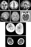
A and B: T2-weighted brain MR images in LAMA2-CMD. Note extensive signal abnormalities of the cerebral white matter while the corpus callosum and the internal capsule are spared (arrows). C: Tl weighted brain MRI in αDG-RD (POMT2).Note thinning of the corpus callosum, the relatively flat pons (arrow) and atrophic and dysplastic cerebellar vermis (arrow head). D and E: T2-weighted MR images in αDG-RD. Note thin corpus callosum, extremely small pons, relatively thick tectum (arrow head), and small and dysplastic cerebellar vermis on the sagittal cut (D). Frontal polymicrogyria (arrow) and abnormal white matter signal is evident on the axial cut (E). F: Tl-weighted MR images in αDG-RD. Note abnormal configuration of the pons and corticospinal tracts and dysplastic cerebellum with cerebellar cysts (arrow) and small vermis (arrow head)). G: Tl-TSE weighted thigh MR images in a COL6-RD, a patient with typical phenotypic UCMD presentation. Note in particular the striated aspect of vastus lateralis caused by outer rim of increased signal (arrow) and increased signal around the central fascia of the rectus femoris (arrow head) (courtesy of Dr. R Carlier). H: Tl-TSE weighted thigh MR images in SEPN1-RM. Note selective involvement of sartorius (arrow), biceps femoris and adductor magnus and sparing of the gracilis (arrow head).

Similar articles

Cited by

  • Urologic and gastrointestinal symptoms in the dystroglycanopathies.
    Crockett CD, Bertrand LA, Cooper CS, Rahhal RM, Liu K, Zimmerman MB, Moore SA, Mathews KD. Crockett CD, et al. Neurology. 2015 Feb 3;84(5):532-9. doi: 10.1212/WNL.0000000000001213. Epub 2015 Jan 7. Neurology. 2015. PMID: 25568299 Free PMC article. Clinical Trial.
  • The genetic basis of undiagnosed muscular dystrophies and myopathies: Results from 504 patients.
    Savarese M, Di Fruscio G, Torella A, Fiorillo C, Magri F, Fanin M, Ruggiero L, Ricci G, Astrea G, Passamano L, Ruggieri A, Ronchi D, Tasca G, D'Amico A, Janssens S, Farina O, Mutarelli M, Marwah VS, Garofalo A, Giugliano T, Sampaolo S, Del Vecchio Blanco F, Esposito G, Piluso G, D'Ambrosio P, Petillo R, Musumeci O, Rodolico C, Messina S, Evilä A, Hackman P, Filosto M, Di Iorio G, Siciliano G, Mora M, Maggi L, Minetti C, Sacconi S, Santoro L, Claes K, Vercelli L, Mongini T, Ricci E, Gualandi F, Tupler R, De Bleecker J, Udd B, Toscano A, Moggio M, Pegoraro E, Bertini E, Mercuri E, Angelini C, Santorelli FM, Politano L, Bruno C, Comi GP, Nigro V. Savarese M, et al. Neurology. 2016 Jul 5;87(1):71-6. doi: 10.1212/WNL.0000000000002800. Epub 2016 Jun 8. Neurology. 2016. PMID: 27281536 Free PMC article.
  • Fukuyama Congenital Muscular Dystrophy.
    Agarwal A, Sabat S, Kanekar S. Agarwal A, et al. Cureus. 2022 Feb 4;14(2):e21902. doi: 10.7759/cureus.21902. eCollection 2022 Feb. Cureus. 2022. PMID: 35273857 Free PMC article.
  • LAMA2 Nonsense Variant in an Italian Greyhound with Congenital Muscular Dystrophy.
    Christen M, Indzhova V, Guo LT, Jagannathan V, Leeb T, Shelton GD, Brocal J. Christen M, et al. Genes (Basel). 2021 Nov 19;12(11):1823. doi: 10.3390/genes12111823. Genes (Basel). 2021. PMID: 34828429 Free PMC article.
  • Clinical utility of multigene analysis in over 25,000 patients with neuromuscular disorders.
    Winder TL, Tan CA, Klemm S, White H, Westbrook JM, Wang JZ, Entezam A, Truty R, Nussbaum RL, McNally EM, Aradhya S. Winder TL, et al. Neurol Genet. 2020 Mar 9;6(2):e412. doi: 10.1212/NXG.0000000000000412. eCollection 2020 Apr. Neurol Genet. 2020. PMID: 32337338 Free PMC article.

References

    1. Nonaka I, Une Y, Ishihara T, Miyoshino S, Nakashima T, Sugita H. A clinical and histological study of Ullrich’s disease (congenital atonic-sclerotic muscular dystrophy) Neuropediatrics. 1981;12:197–208. - PubMed
    1. Kobayashi O, Hayashi Y, Arahata K, Ozawa E, Nonaka I. Congenital muscular dystrophy: clinical and pathologic study of 50 patients with the classical (Occidental) merosin-positive form. Neurology. 1996;46:815–818. - PubMed
    1. Reed UC. Congenital muscular dystrophy. Part I: a review of phenotypical and diagnostic aspects. Arq Neuropsiquiatr. 2009;67:144–168. - PubMed
    1. Reed UC. Congenital muscular dystrophy. Part II: a review of pathogenesis and therapeutic perspectives. Arq Neuropsiquiatr. 2009;67:343–362. - PubMed
    1. Mercuri E, Muntoni F. The ever-expanding spectrum of congenital muscular dystrophies. Ann Neurol. 2012;72:9–17. - PubMed

Publication types