. 2014 Apr 8;22(4):515-525.
doi: 10.1016/j.str.2014.01.010. Epub 2014 Feb 27.

(19)F NMR reveals multiple conformations at the dimer interface of the nonstructural protein 1 effector domain from influenza A virus

Affiliations

(19)F NMR reveals multiple conformations at the dimer interface of the nonstructural protein 1 effector domain from influenza A virus

James M Aramini et al. Structure. .

Abstract

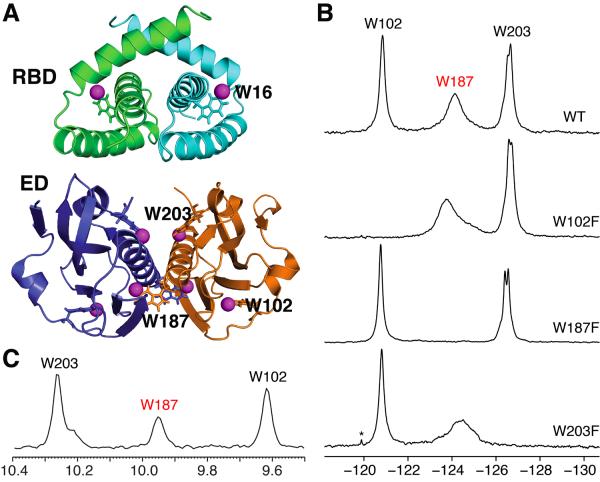
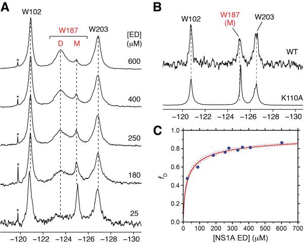
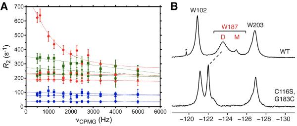
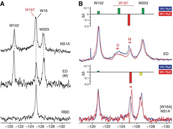
Nonstructural protein 1 of influenza A virus (NS1A) is a conserved virulence factor comprised of an N-terminal double-stranded RNA (dsRNA)-binding domain and a multifunctional C-terminal effector domain (ED), each of which can independently form symmetric homodimers. Here we apply (19)F NMR to NS1A from influenza A/Udorn/307/1972 virus (H3N2) labeled with 5-fluorotryptophan, and we demonstrate that the (19)F signal of Trp187 is a sensitive, direct monitor of the ED helix:helix dimer interface. (19)F relaxation dispersion data reveal the presence of conformational dynamics within this functionally important protein:protein interface, whose rate is more than three orders of magnitude faster than the kinetics of ED dimerization. (19)F NMR also affords direct spectroscopic evidence that Trp187, which mediates intermolecular ED:ED interactions required for cooperative dsRNA binding, is solvent exposed in full-length NS1A at concentrations below aggregation. These results have important implications for the diverse roles of this NS1A epitope during influenza virus infection.


Figures

Figure 1
Figure 1
(A) Locations of the 5-F-Trp residues in the dimer structures of Ud NS1A domains. Top, Ud NS1A RBD (PDB ID, 1NS1) (Chien et al., 1997). Bottom, Ud NS1A ED (PDB ID, 3EE9) (Xia et al., 2009). The fluorine atoms are shown as magenta spheres, and the tryptophan residues within one subunit of each dimer are labeled. Structures were rendered using PyMOL (PyMOL Molecular Graphics System, Version 1.4 Schrödinger, LLC). (B) 19F NMR spectra of 5-F-Trp labeled Ud NS1A ED (530 μM), and its W102F (400 μM), W187F (400 μM), and W203F (200 μM) mutants in high salt pH 8 buffer. Asterisks denote trace amounts of protease inhibitors remaining after purification. Note that the splitting observed for Trp203 under these conditions is more pronounced for the W187F mutant which is a monomer in solution (data not shown). (C) Projection of the indole amide region along the 1H dimension from an 800 MHz 2D 1H-15N TROSY-HSQC spectrum of 400 μM 15N-enriched Ud NS1A ED in pH 6.9 buffer at 27 °C (Aramini et al., 2011).
Figure 2
Figure 2
19F NMR is a direct probe of Ud NS1A ED homodimerization. (A) Concentration dependence of the 19F NMR signal of Trp187 within 5-F-Trp labeled Ud NS1A ED in low salt pH 8 buffer. Resonances corresponding to Trp187 in the dimer and monomer states are denoted by `D' and `M', respectively. Asterisks denote trace amounts of residual protease inhibitors after purification. (B) 19F NMR spectra of 5-F-Trp labeled monomeric 25 μM Ud NS1A ED (top) and 500 μM [K110A] NS1A ED (bottom), a known monomeric effector domain mutant (Aramini et al., 2011), in high salt pH 8 buffer. (C) Fit of the fraction of 19F dimer resonance volume as a function of total Ud NS1A ED concentration. Dashed lines represent 95% confidence bounds for the fit.
Figure 3
Figure 3
19F NMR CPMG relaxation dispersion data for Trp102 (squares), Trp187 (circles), and Trp203 (triangles) in 5-F-Trp labeled 600 μM Ud NS1A ED (red), 200 μM [K110A] NS1A ED (blue), and 500 μM[C116S,G183C] NS1A ED (disulfide form; no DTT) (green) in low salt pH 8 buffer at 20 °C. (B) 19F NMR spectra of 5-F-Trp labeled Ud NS1A ED (top) and disulfide-bonded 500 μM [C116S,G183C] NS1A ED (bottom), in low salt pH 8 buffer. The downfield shift of the 19F resonance for Trp187 is indicated by a dashed line. The native Cys116 was mutated to a serine in order to prevent a potential mixture of disulfide adducts.
Figure 4
Figure 4
Field dependent 19F CPMG relaxation dispersion data for Trp102 (squares), Trp187 (circles), and Trp203 (triangles) of 5-F-Trp labeled Ud NS1A ED dimer (600 μM) in low salt pH 8 buffer. Transverse relaxation rates (R2) as a function of νCPMG acquired at magnetic field strengths of 11.7 T (470.18 MHz) and 14.1 T (564.62 MHz) are shown in red and cyan, respectively. The relaxation dispersion curves for Trp187 at both fields were fit to a two-state exchange model (Carver and Richards, 1972),yielding the following results: kex = 6430 ± 1180 s−1; R20 (470.18 MHz) = 269 ± 18 s−1; R20 (564.62 MHz) = 328 ± 16 s−1; α = 2.3 ± 0.6.
Figure 5
Figure 5
(A) 19F NMR spectra of 5-F-Trp labeled Ud NS1A′ (50 μM), monomeric ED (25 μM), and RBD (200 μM) in high salt pH 8 buffer. (B) Overlay of 19F NMR spectra of 5-F-Trp labeled Ud NS1A ED (top) and [W16A] NS1A′ (bottom) in 10% (blue) and 90% (red)2H2O. Sample conditions: i) 150μM (10% 2H2O) and 300 μM (90% 2H2O) NS1A ED in low salt pH 8 buffer; ii) 35 μM [W16A] NS1A′ in high salt pH 8 buffer. Insets above both spectral overlays depict the change in 19F chemical shift (Δδ) and degree of solvent exposure for each 5-F-Trp colored as follows: green, buried, red, exposed, and yellow, partially exposed. Resonances corresponding to Trp187 in the dimer and monomer states of NS1A ED are denoted by `D' and `M', respectively.
Figure 6
Figure 6
Superimposed crystal structures of NS1A ED showing “snapshots” of the conformational heterogeneity of the helix:helix interface. All dimer structures were oriented to optimally-superimpose the indole ring atoms of Trp187 (shown in grey). (A) View showing the range of movements of Trp187′ in the second protomer as well as the long helix (residues 171 to 188) in both protomers (α1 and α1′). Crystallographic dimers are colored based on the relative angle between Trp187 and Trp187′ indole rings, from smallest (yellow) to largest (blue). (B) Close-up view of the environment that would be sensed by a fluorine atom at the C5 position of Trp187 (magenta sphere). Residues in the second protomer that are consistently within 5 Å of the fluorine position in Trp187 of the first protomer are labeled. Structure coordinates correspond to selected crystallographic dimer units from the NS1A ED literature coordinates for the following influenza A strains and crystal forms: A/Puerto Rico/8/1934 (H1N1), PDB ID, 2GX9 (Bornholdt and Prasad, 2006); A/Udorn/307/1972 (H3N2), PDB IDs, 3EE8 and 3EE9 (Xia et al., 2009); A/California/07/2009 (H1N1), PDB ID, 3M5R; A/Reassortant/IVR108 (Sydney/5/1995 × Puerto Rico/8/1934) (H3N2), PDB ID, 3O9U (Kerry et al., 2011). Structures were rendered using PyMOL.

Similar articles

Cited by

References

    1. Acchione M, Lee YC, DeSantis ME, Lipschultz CA, Wlodawer A, Li M, Shanmuganathan A, Walter RL, Smith-Gill S, Barchi JJ., Jr. Specific fluorine labeling of the HyHEL10 antibody affects antigen binding and dynamics. Biochemistry. 2012;51:6017–6027. - PMC - PubMed
    1. Acton TB, Xiao R, Anderson S, Aramini J, Buchwald WA, Ciccosanti C, Conover K, Everett J, Hamilton K, Huang YJ, et al. Preparation of protein samples for NMR structure, function, and small-molecule screening studies. Methods Enzymol. 2011;493:21–60. - PMC - PubMed
    1. Ahmed AH, Loh AP, Jane DE, Oswald RE. Dynamics of the S1S2 glutamate binding domain of GluR2 measured using 19F NMR spectroscopy. J Biol Chem. 2007;282:12773–12784. - PubMed
    1. Aramini JM, Ma LC, Zhou L, Schauder CM, Hamilton K, Amer BR, Mack TR, Lee HW, Ciccosanti CT, Zhao L, et al. Dimer interface of the effector domain of non-structural protein 1 from influenza A virus: an interface with multiple functions. J Biol Chem. 2011;286:26050–26060. - PMC - PubMed
    1. Ayllon J, Russell RJ, Garcia-Sastre A, Hale BG. Contribution of NS1 Effector Domain Dimerization to Influenza A Virus Replication and Virulence. J Virol. 2012;86:13095–13098. - PMC - PubMed

Publication types

MeSH terms

LinkOut - more resources