Review
. 2014 Apr;22(4):183-91.
doi: 10.1016/j.tim.2014.01.010. Epub 2014 Feb 26.

Bat-derived influenza-like viruses H17N10 and H18N11

Affiliations
Review

Bat-derived influenza-like viruses H17N10 and H18N11

Ying Wu et al. Trends Microbiol. 2014 Apr.

Abstract

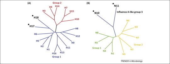
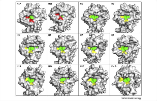
Shorebirds and waterfowls are believed to be the reservoir hosts for influenza viruses, whereas swine putatively act as mixing vessels. The recent identification of two influenza-like virus genomes (designated H17N10 and H18N11) from bats has challenged this notion. A crucial question concerns the role bats might play in influenza virus ecology. Structural and functional studies of the two major surface envelope proteins, hemagglutinin (HA) and neuraminidase (NA), demonstrate that neither has canonical HA or NA functions found in influenza viruses. However, putative functional modules and domains in other encoded proteins are conserved, and the N-terminal domain of the H17N10 polymerase subunit PA has a classical structure and function. Therefore, potential genomic reassortments of such influenza-like viruses with canonical influenza viruses cannot be excluded at this point and should be assessed.

Keywords: H17N10; H18N11; PA; bat-derived influenza-like virus; hemagglutinin (HA); neuraminidase (NA); reassortment.


Figures

Figure 1
Figure 1
Phylogenetic trees of the hemagglutinin (HA) and neuraminidase (NA) genes of all known influenza A virus subtypes including H17/18 and NA-like N10/11. (A) The categorized HA molecules can be grouped into two groups, group 1 and group 2. The bat-derived H17 and H18 (each marked with a star) belong to group 1, displaying a typical sequence feature of group 1 HA molecules. (B) All the known NAs could also be grouped into two groups: group 1 and group 2. The bat-derived N10 and N11 (each marked with a star) do not belong to either group. They are NA homologs or could be called NA-like molecules. Here we propose that they be categorized as influenza A-like group 3.
Figure 2
Figure 2
Configurations of avian and human receptor analogs. (A) The avian receptor analog. The Sia-1 (sialic acid, SA) is linked to Gal-2 (galactose) via an α2,3-glycosidic bond. The glycans extend forward and the hydrophilic glycosidic oxygen atom is exposed to the receptor binding site. (B) The human receptor analog. The Sia-1 is linked to Gal-2 via an α2,6-glycosidic bond. The glycans fold back and the hydrophobic C6 atom is exposed to the receptor binding site. In both (A) and (B) N-acetylglucosamine is abbreviated as GlcNAc-3; black arrows indicate the orientations of the glycosidic linkage.
Figure 3
Figure 3
Receptor-binding ‘head’ domains of all the hemagglutinins (HAs) with known structures (including influenza A and B) showing the SA binding sites. This figure clearly shows that H17 and H18 have a smaller SA-binding cavity – with two acidic amino acids (labeled in red) in comparison to the non-charged amino acids in all the other HA molecules (labeled in yellow). Note, the base of the receptor binding groove is labeled in green; both H17 and H18 show a ‘closed’ conformation, and that of H18 is even smaller.
Figure 4
Figure 4
Enzymatic active sites and the adjacent 150-loop structures of all the neuraminidases (NAs) with known structures (including influenza B virus) and NA-like N10 and N11. All the NA structures have been crystallographically resolved except for N7. Amino acids of the 150-loop are labeled in yellow and the substrate-binding groove is labeled in blue. Members from group 1 have a 150-cavity (except for N1 of 2009 pH1N1), which group 2 members otherwise lack. N10 and N11 also lack a 150-cavity, and have a more open structure which does not support SA binding. Under some circumstances, 09N1 can open (I223R mutation) and N2 can half-open (oseltamivir carboxylate-induced). After binding to the inhibitors, group 1 members usually close their 150-cavity (N5 is shown with oseltamivir bound).
Figure 5
Figure 5
Schematic of the 14 influenza A virus-encoded proteins showing the functional and structural modules or domains. All the known modules are indicated and are highly conserved in the genomes of both H17N10 and H18N11. Abbreviations: ‘?’, unknown functions; CT, cytoplasmic tail; MT, mitochondrial targeting sequence; NES, nuclear export signal; NLS, nuclear localization signal; PBS, promoter binding site; RdRp, RNA-dependent RNA polymerase; RNP, ribonucleoprotein; TM, transmembrane anchor. Amino acid numbering is based on all the protein sequences of the 1918 pandemic H1N1 virus [Genbank: ABA55038.1(PB2), ABA55039.1(PB1/PB1N40), ABW36320.1(PB1-F2), ABA55040.1(PA), AGG82783.1(PA-X), AAD17229.1(HA), AAV48837.1(NP), AAF77036.1(NA), AAN06597.1(M1), AAN06598.1(M2/M42), AAK14368.1(NS1), AAK14369.1(NS2)].

References

    1. Palese P., Shaw M.L. Orthomyxoviridae: the viruses and their replication. In: Knipe D.M., Howley P.M., editors. Fields Virology. 4th edn. Lippincott Williams & Wilkins; 2007. pp. 1647–1690.
    1. Cox N.J., Subbarao K. Global epidemiology of influenza: past and present. Annu. Rev. Med. 2000;51:407–421. - PubMed
    1. Kobasa D. Enhanced virulence of influenza A viruses with the haemagglutinin of the 1918 pandemic virus. Nature. 2004;431:703–707. - PubMed
    1. Novel Swine-Origin Influenza A (H1N1) Virus Investigation Team Emergence of a novel swine-origin influenza A (H1N1) virus in humans. N. Engl. J. Med. 2009;360:2605–2615. - PubMed
    1. Webster R.G. Evolution and ecology of influenza A viruses. Microbiol. Rev. 1992;56:152–179. - PMC - PubMed

Publication types