

Micropattern width dependent sarcomere development in human ESC-derived cardiomyocytes
- PMID: 24582552
- PMCID: PMC4026015
- DOI: 10.1016/j.biomaterials.2014.02.001
Micropattern width dependent sarcomere development in human ESC-derived cardiomyocytes
Abstract
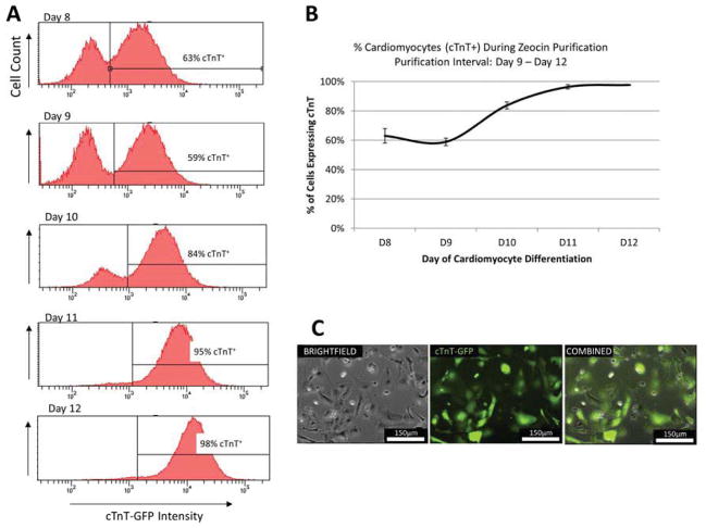
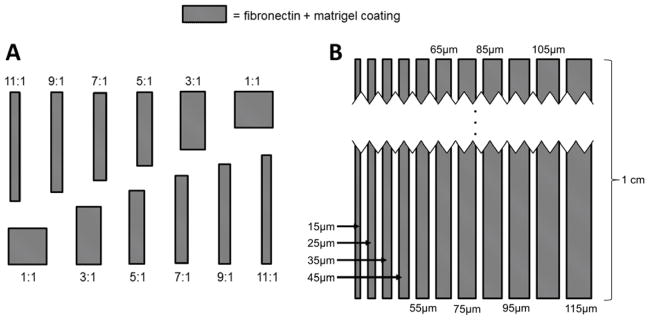
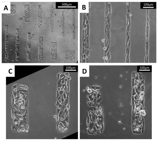
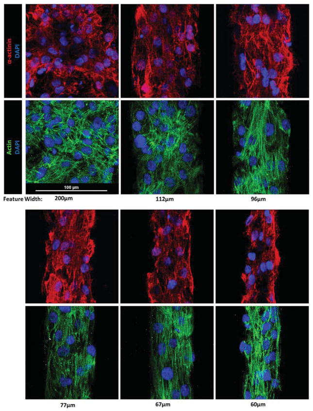
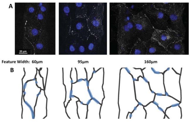
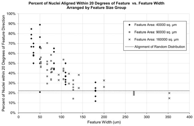
In this study, human embryonic stem cell-derived cardiomyocytes were seeded onto controlled two-dimensional micropatterned features, and an improvement in sarcomere formation and cell alignment was observed in specific feature geometries. High-resolution photolithography techniques and microcontact printing were utilized to produce features of various rectangular geometries, with areas ranging from 2500 μm(2) to 160,000 μm(2). The microcontact printing method was used to pattern non-adherent poly(ethylene glycol) regions on gold coated glass slides. Matrigel and fibronectin extracellular matrix (ECM) proteins were layered onto the gold-coated glass slides, providing a controlled geometry for cell adhesion. We used small molecule-based differentiation and an antibiotic purification step to produce a pure population of immature cardiomyocytes from H9 human embryonic stem cells (hESCs). We then seeded this pure population of human cardiomyocytes onto the micropatterned features of various sizes and observed how the cardiomyocytes remodeled their myofilament structure in response to the feature geometries. Immunofluorescence was used to measure α-actinin expression, and phalloidin stains were used to detect actin presence in the patterned cells. Analysis of nuclear alignment was also used to determine how cell direction was influenced by the features. The seeded cells showed clear alignment with the features, dependent on the width rather than the overall aspect ratio of the features. It was determined that features with widths between 30 μm and 80 μm promoted highly aligned cardiomyocytes with a dramatic increase in sarcomere alignment relative to the long axis of the pattern. This creation of highly-aligned cell aggregates with robust sarcomere structures holds great potential in advancing cell-based pharmacological studies, and will help researchers to understand the means by which ECM geometries can affect myofilament structure and maturation in hESC-derived cardiomyocytes.
Keywords: Cardiac tissue engineering; Cardiomyocyte; Cell morphology; Micropatterning; Stem cell; Surface modification.
Copyright © 2014 Elsevier Ltd. All rights reserved.
Figures

Similar articles
-
Tissue-engineered cardiac patch for advanced functional maturation of human ESC-derived cardiomyocytes.Biomaterials. 2013 Jul;34(23):5813-20. doi: 10.1016/j.biomaterials.2013.04.026. Epub 2013 May 2. Biomaterials. 2013. PMID: 23642535 Free PMC article.
-
Engineering a functional three-dimensional human cardiac tissue model for drug toxicity screening.Biofabrication. 2017 May 11;9(2):025011. doi: 10.1088/1758-5090/aa6c3a. Biofabrication. 2017. PMID: 28393762
-
Cell Architecture and Dynamics of Human Induced Pluripotent Stem Cell-Derived Cardiomyocytes (hiPSC-CMs) on Hydrogels with Spatially Patterned Laminin and N-Cadherin.ACS Appl Mater Interfaces. 2025 Jan 8;17(1):174-186. doi: 10.1021/acsami.4c11934. Epub 2024 Dec 16. ACS Appl Mater Interfaces. 2025. PMID: 39680735 Free PMC article.
-
Human pluripotent stem cell-derived cardiomyocytes for heart regeneration, drug discovery and disease modeling: from the genetic, epigenetic, and tissue modeling perspectives.Stem Cell Res Ther. 2013 Aug 14;4(4):97. doi: 10.1186/scrt308. Stem Cell Res Ther. 2013. PMID: 23953772 Free PMC article. Review.
-
Sarcomere maturation: function acquisition, molecular mechanism, and interplay with other organelles.Philos Trans R Soc Lond B Biol Sci. 2022 Nov 21;377(1864):20210325. doi: 10.1098/rstb.2021.0325. Epub 2022 Oct 3. Philos Trans R Soc Lond B Biol Sci. 2022. PMID: 36189811 Free PMC article. Review.
Cited by
-
Bioengineering approaches to mature induced pluripotent stem cell-derived atrial cardiomyocytes to model atrial fibrillation.Exp Biol Med (Maywood). 2021 Aug;246(16):1816-1828. doi: 10.1177/15353702211009146. Epub 2021 Apr 25. Exp Biol Med (Maywood). 2021. PMID: 33899540 Free PMC article. Review.
-
Designing Biomaterial Platforms for Cardiac Tissue and Disease Modeling.Adv Nanobiomed Res. 2021 Jan;1(1):2000022. doi: 10.1002/anbr.202000022. Epub 2020 Oct 16. Adv Nanobiomed Res. 2021. PMID: 33709087 Free PMC article.
-
Bioresorbable Polymeric Scaffold in Cardiovascular Applications.Int J Mol Sci. 2020 May 13;21(10):3444. doi: 10.3390/ijms21103444. Int J Mol Sci. 2020. PMID: 32414114 Free PMC article. Review.
-
Influence of Micropatterning on Human Periodontal Ligament Cells' Behavior.Biophys J. 2018 Apr 24;114(8):1988-2000. doi: 10.1016/j.bpj.2018.02.041. Biophys J. 2018. PMID: 29694875 Free PMC article.
-
In Vitro Microscale Models for Embryogenesis.Adv Biosyst. 2018 Jun;2(6):1700235. doi: 10.1002/adbi.201700235. Epub 2018 May 7. Adv Biosyst. 2018. PMID: 30533517 Free PMC article.
References
-
- Xu J, Kochanek KD, Murphy S, Tejada-Vera B. Deaths: final data for 2007. Hyattsville, MD: National Center for Health Statistics; - PubMed
- Natl Vital Stat Rep. 2010;58:1–135.
-
- Kattman SJ, Witty AD, Gagliardi M, Dubois NC, Niapour M, Hotta A, et al. Stage-specific optimization of activin/nodal and BMP signaling promotes cardiac differentiation of mouse and human pluripotent stem cell lines. Cell Stem Cell. 2011;8(2):228–240. - PubMed
-
- Laflamme MA. Cardiomyocytes derived from human embryonic stem cells in pro-survival factors enhance function of infarcted rat hearts. Nat Biotechnol. 2007;25:1015–1024. - PubMed
Publication types
MeSH terms
Substances
Grants and funding
LinkOut - more resources
Full Text Sources
Other Literature Sources
