. 2014 Jun:81:274-82.
doi: 10.1016/j.neuropharm.2014.02.012. Epub 2014 Feb 26.

Modulation of HCN channels in lateral septum by nicotine

Affiliations

Modulation of HCN channels in lateral septum by nicotine

Sodikdjon A Kodirov et al. Neuropharmacology. 2014 Jun.

Abstract

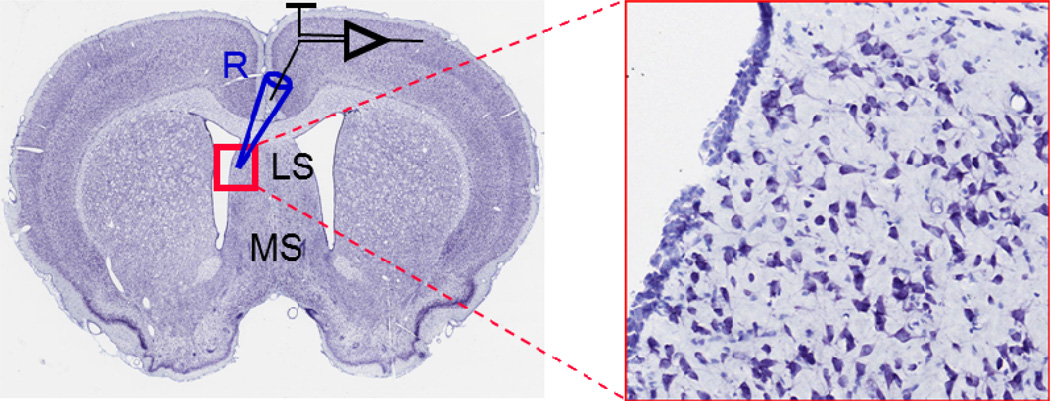
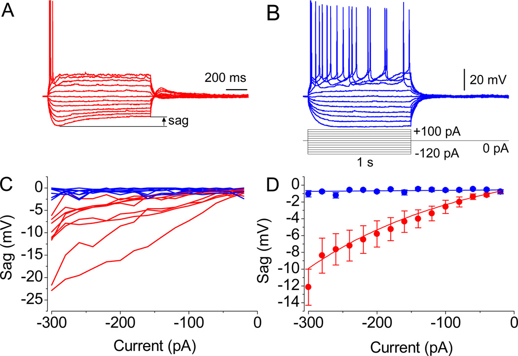
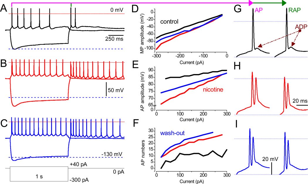
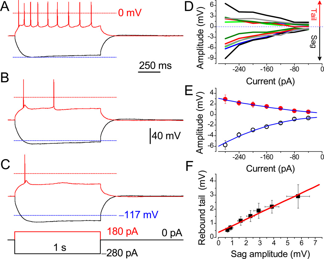
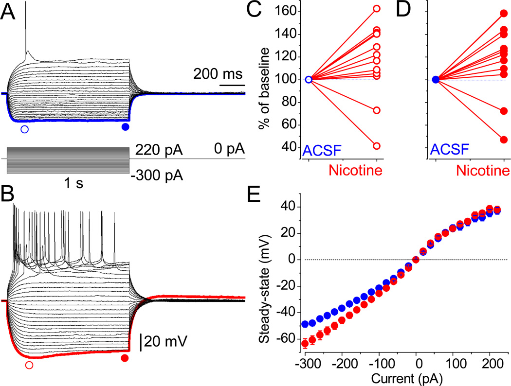
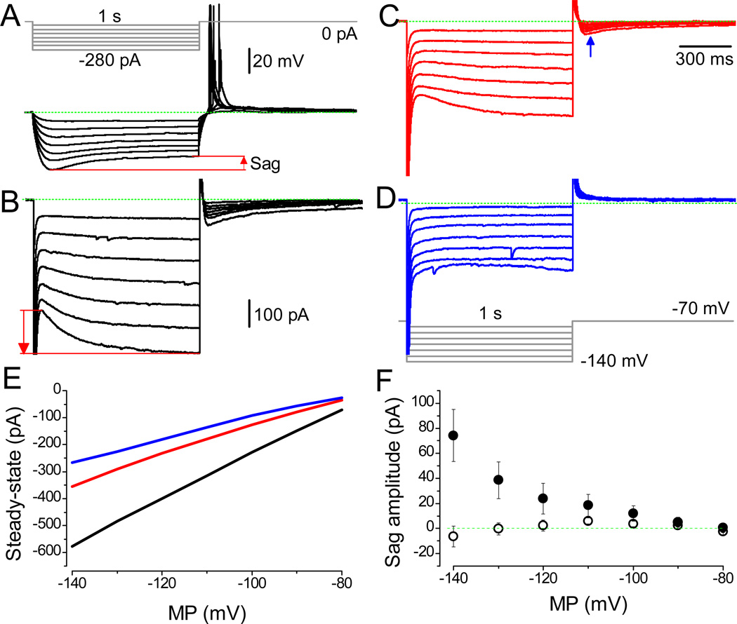
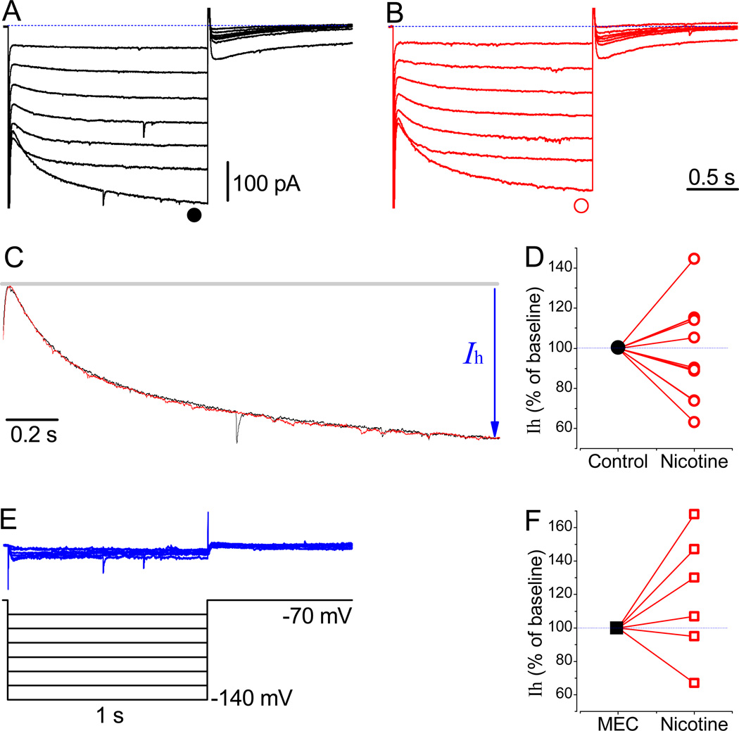
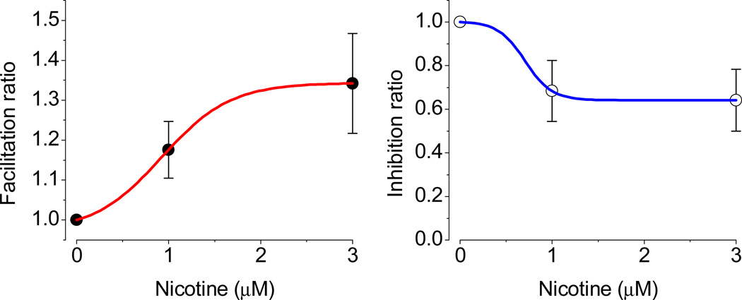
The effects of addictive drugs most commonly occur via interactions with target receptors. The same is true of nicotine and its multiple receptors in a variety of cell types. However, there are also side effects for given substances that can dramatically change cellular, tissue, organ, and organism functions. In this study, we present evidence that nicotine possesses such properties, and modulates neuronal excitability. We recorded whole-cell voltages and currents in neurons situated in the dorsal portion of the lateral septum in acute coronal brain slices of adult rats. Our experiments in the lateral septum revealed that nicotine directly affects HCN - hyperpolarization-activated cyclic nucleotide gated non-selective cation channels. We demonstrate that nicotine effects persist despite the concurrent application of nicotinic acetylcholine receptors' antagonists - mecamylamine, methyllycaconitine, and dihydro-β-erythroidine. These results are novel in regard to HCN channels in the septum, in general, and in their sensitivity to nicotine, in particular.

Keywords: Action potentials; HCN channels; Sag potential; Septal neurons; h-Current.


Figures

Fig. 1
Fig. 1
Coronal slice from rat brain and location of recorded neurons at higher magnification. R – recording electrode, LS – lateral septum, MS – medial septum. Modified from www.brainmaps.org (Mikula et al., 2007).
Fig. 2
Fig. 2
Action potential waveforms. (A and B) Evoked APs in two different neurons. Note the significant sag in MP in A and its absence in B, as revealed upon the injection of currents of relatively low amplitude. (C) Representation of sag’s magnitude from all recorded neurons. (D) The mean amplitude values in cells with properties resembling the one shown in A (n = 9, P < 0.01 at −300 to −40 pA, paired t test) and B (n = 9, P = 0.5). Resting membrane potential was −55.2 mV in A and −52.2 mV in B. Identical scale bars are shown.
Fig. 3
Fig. 3
Nicotine facilitates neuronal excitably in LS. (A–C) Recordings were obtained from the same neuron in the absence and the presence of 3 µM nicotine and upon wash-out. The waveforms of MP during depolarizing (40 pA) and hyperpolarizing (−300 pA) current steps are superimposed. The red and blue dashed lines mark MP at ±0 mV and −130 mV, respectively. (D–F) Values before (black traces) and after the application of 3 µM nicotine (red) and upon the wash-out (blue). The analyzed parameters are: (D) the peak MP response to hyperpolarizing currents ranging from −20 pA to −300 pA in 20-pA increments; (E) the amplitude of the first AP upon depolarization by current steps (20–300 pA); (F) the number of APs per second. Note that, under control conditions, no AP was triggered at 20 pA (black). (G–I) The phenotype of both AP (left) and RAP (right) in a train, in the absence (black) or presence of 3 µM nicotine (red), and during wash-out (blue). Note the transition of a single AP with prominent ADP (under control conditions) into double spikes in the presence of nicotine. Scale bars for A–C and G–I are identical.
Fig. 4
Fig. 4
Suppression of evoked action potentials by nicotine in a subset of neurons. APs were evoked in LS neuron before (A) and after (B) treatment with 3 µM nicotine, and after 30 min wash-out (C). The APs were evoked from the RMP of −60.1 mV under identical conditions. Lower dashed lines are MP at −117 mV. Identical scale bars apply for A–C. (D) The sag and rebound tail potentials in different neurons are color-matched. (E) Mean amplitudes of rebound tail (●) and sag potentials (○, n = 6). Note that the amplitude of sag is two-fold greater than the tail potential. (F) The linear correlation between values of sag and tail potentials (R = 0.99).
Fig. 5
Fig. 5
Nicotine-mediated sag. (A) The MP in neurons of the LS was recorded in response to hyperpolarizing currents under identical conditions, as in Figure 2. (B) Within 15 min of exposure to nicotine, sag and related rebound tail potentials appear. Scale bars for A and B are identical. The blue and red symbols indicate the points taken for statistical analyses. (C and D) The individually normalized peak and steady-state amplitude values for all tested neurons. (E) The mean amplitude values of the steady-state potentials were increased by nicotine only at more hyperpolarized MP (for example, at −200 pA: −45.8 ± 2.4 mV vs. −36.8 ± 1.7 mV, n = 10, P = 0.001). Error bars are very small under control conditions.
Fig. 6
Fig. 6
Effects of nicotine under nAChRs blockade. (A) Increase in sag and rebound tail potentials by nicotine despite the presence of antagonist, DHβE. Note the spontaneous postsynaptic potentials under control conditions. (B) Amplitude values obtained in the presence of 100 nM DHβE (●, n = 9). The presented data subdivide into two groups reflecting nicotine’s dual effects: an increase (○, n = 6) and a decrease (○, n = 3) in peak amplitudes. (C) Under these conditions, values of pooled data for all neurons were not different in DHβE (●) and nicotine (○) sets of experiments (n = 9). (D–F) The individually normalized values for each cell reveal the same tendency in the presence of either DHβE (n = 9), MLA (1 µM; n = 7), or both (n = 7). Note that only one cell (○) in E was not affected by nicotine in the presence of MLA. Scale bars are shown.
Fig. 7
Fig. 7
Selective inhibition of HCN channel mediated non-selective cationic currents. (A) The typical and comparable response of an LS neuron. Note RAPs upon termination of hyperpolarization; RMP: −63.9 mV. (B) Activation of Ih from the holding potential of −70 mV by the pulse protocol shown in D. The Ih, i.e. the counterpart of the sag potential, was observed readily at more hyperpolarized test potentials. (C and D) Dose-dependent decrease in amplitude by 1 and 10 µM ZD 7288, respectively. (E) The application of ZD 7288 affects the steady–state amplitudes. The Ih are shown under control conditions (black line) and in the presence of 1 µM (red) and 10 µM ZD 7288 (blue). (F) Consistently, the two groups confirming the absence (○, n = 4) and the presence (●, n = 8) of HCN-mediated currents are distinguished. The amplitude’s scale bar for A is shown, while the time is reflected by the pulse protocol. Scale bars indicating current amplitude are identical for B–D.
Fig. 8
Fig. 8
Effects of nicotine and MEC on Ih. (A) The Ih recorded at a holding potential of −70 mV using the protocol shown in E. (B) Traces are recorded 15 min after the application of 3 µM nicotine. (C) Normalized and superimposed traces revealing the similar activation kinetics of HCN channels in the absence of and the presence of nicotine at −140 mV, respectively. (D) Normalized Ih at −130 mV (●, n = 11). (E) The subtracted nicotine-sensitive currents. (F) Normalized Ih in the presence of a non-selective inhibitor of nAChRs, 10 µM MEC (■, n = 6). The time and amplitude scale bars are identical for A, B, and E.
Fig. 9
Fig. 9
Cumulative dose response of nicotine in LS neurons. The facilitation and inhibition are shown for subgroups of neurons. Under identical experimental conditions, the facilitation saturated at 3 µM, while inhibition was already saturated at 1 µM (n = 4). The EC50 for facilitation was 0.92 µM (n = 6).

Similar articles

Cited by

References

    1. Altomare C, Bucchi A, Camatini E, Baruscotti M, Viscomi C, Moroni A, DiFrancesco D. Integrated allosteric model of voltage gating of HCN channels. J Gen Physiol. 2001;117:519–532. - PMC - PubMed
    1. Araque A, Cattaert D, Buno W. Cd2+ regulation of the hyperpolarization-activated current IAB in crayfish muscle. J Gen Physiol. 1995;105:725–744. - PMC - PubMed
    1. Bland BH, Colom LV. Extrinsic and intrinsic properties underlying oscillation and synchrony in limbic cortex. Prog Neurobiol. 1993;41:157–208. - PubMed
    1. Brager DH, Lewis AS, Chetkovich DM, Johnston D. Short- and long-term plasticity in CA1 neurons from mice lacking h-channel auxiliary subunit TRIP8b. J Neurophysiol. 2013;110:2350–2357. - PMC - PubMed
    1. Brown H, Difrancesco D, Noble S. Cardiac pacemaker oscillation and its modulation by autonomic transmitters. J Exp Biol. 1979;81:175–204. - PubMed

Publication types